By Heloa | 30 April 2025

Infant hemangioma: everything parents need to know

8 minutes
de lecture
angiome-bébé-illustration-malo

By Heloa | 30 April 2025

Infant hemangioma: everything parents need to know

8 minutes
angiome-bébé-illustration-malo

Par Heloa, le 30 April 2025

Infant hemangioma: everything parents need to know

8 minutes
de lecture
angiome-bébé-illustration-malo

The birth of a baby ignites countless questions, but nothing grabs attention quite like a bright red or purplish mark blossoming on fragile skin. That first glimpse—a splash of colour, a bump rising where yesterday there was none—can be startling. Is it painful? Will it grow larger? Could it harm my child? Such worries are common when infant hemangioma makes its entrance, often called a “strawberry mark” for its vivid hue and texture. Many parents find themselves scanning their child for changes, juggling between reassurance and concern. What is certain: knowing what to expect, understanding treatments, and recognising risks helps families move from uncertainty to confident care.

Explore how infant hemangioma emerges, why it chooses some babies, how it evolves, and most importantly, how parents can protect, soothe, and support a child as these vascular tumours chart their unpredictable journey—whether fading quietly with time or demanding medical attention.

What Is Infant Hemangioma? Recognising the Mark and Its Nature

A small, soft growth, strikingly red or at times bluish, suddenly appears—barely noticeable at first, but then it starts to expand. Infant hemangioma stands as the most prevalent benign vascular tumour in infancy, appearing in about 4–5% of babies. Far from being a rarity, it’s seen more frequently in girls, premature infants, and babies of lower birth weight, sometimes clustering along the face or scalp, or surprising parents on the trunk or limbs.

Structurally, an infant hemangioma is made up of an overgrowth of delicate blood vessels (capillaries), forming just under or on the skin. Think of it as a tangle of vessels working overtime—unusually active, but not cancerous. Its drama lies in three main acts: first, rapid growth (the proliferative phase, most intense within 3 to 6 months); second, a stable plateau where size steadies; and third, the gradual fading (the involution phase) which may take several years, often leaving behind a faint shadow, a slight change in texture, perhaps a silvery patch.

How is this different from other marks? Unlike port wine stains or vascular malformations, which stay the same over time, an infant hemangioma has a unique life cycle—intense change, then retreat. One clue for doctors: the GLUT-1 protein marker, present only in these particular tumours.

Why Do Infant Hemangiomas Develop? Unravelling the Risk Factors

Curiosity runs high: Why my child? The answer isn’t straightforward. The precise triggers behind infant hemangioma remain partly mysterious, but science points to a web of influences:

  • Prematurity and low birth weight: The smallest and most fragile babies are at higher risk.
  • Female gender: Girls, by a margin of up to five to one, outnumber boys.
  • Maternal and pregnancy factors: Ageing mothers, twins or triplets, and certain pregnancy procedures (like chorionic villus sampling).
  • Hypoxic stress: Episodes of lower oxygen during fetal life may send a signal—blood vessels, grow! This fires up angiogenic factors (proteins like VEGF and GLUT-1).
  • Genetic and syndromic cues: Sometimes, hemangiomas cluster in families, or announce themselves as part of syndromes like PHACE—where other organs such as the heart or brain may also show abnormalities.

For most, however, there is no clear family link—just a sporadic quirk of development.

The Making of an Infant Hemangioma: Science Under the Skin

Cells with big ambition—endothelial progenitor cells—are at the core. These are the architects of new blood vessels, usually keeping growth in check. In infant hemangioma, this control slips. Some experts talk of the “placental provenance theory”—a fascinating idea that cells with traits like those from the placenta hitch a ride to the skin, starting a chain reaction. Their signal? Growth factors such as VEGF, triggering new vessels relentlessly.

Visualise the progression:

  • Early days: A pale patch, almost invisible, or a faint hint of pink.
  • Rapid ascent: Within weeks, a mushrooming of colour—red, raised, sometimes lobulated—heralds the proliferative burst.
  • Deep components: If the activity is beneath the surface, bluish swelling emerges—softer, almost sponge-like.
  • Retreat: By toddlerhood, slow shrinking sets in, sometimes leaving fibrofatty tissue or delicate telangiectasia (tiny red threads).

Markers such as VEGF, CD31, and especially GLUT-1 help unravel the tumour’s true nature for pathologists, making distinction from other vascular lesions possible.

How Infant Hemangiomas Look, Grow, and Change

Each hemangioma writes its own story, but some themes repeat:

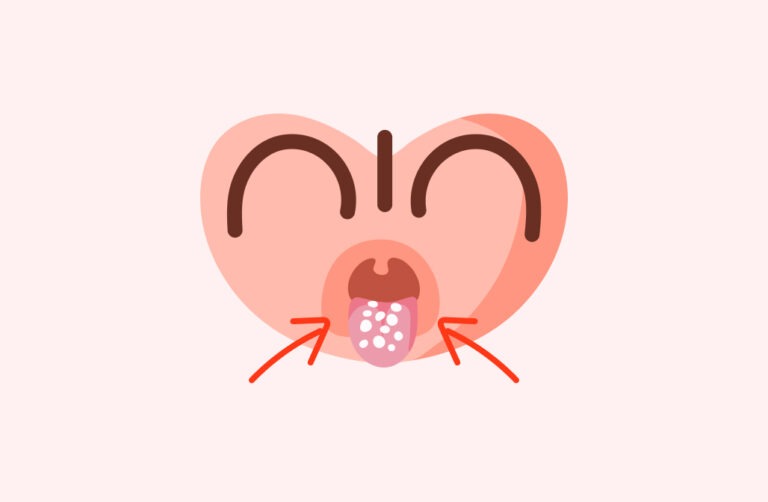
  • Superficial hemangioma: Like a strawberry perched on the skin—bright red, defined, and often alarming in its speed.
  • Deep hemangioma: Hiding below, it may look bluish or purplish, emerging as a soft, compressible mass.
  • Mixed hemangioma: A blend of both, adding complexity to colour and shape.

Where do they appear? While face and neck lead (especially eyelids, lips, or scalp), they can also sprout on limbs, torso, or in rare instances, within internal organs such as the liver. A focal lesion remains small and defined, a segmental one stretches along a larger developmental pathway, while multifocal types sprinkle several sites.

What should parents watch for? Early expansion is common—expect a growth spurt in the first few months, then a calming phase, and ultimately a slow, patient regression.

Typical Locations and Why They Matter

Location matters. A vivid infant hemangioma on the cheek or eyelid commands more worry than a barely visible patch on the leg. Most often, these vascular marks nestle on visible territory: head, neck, eyelids, hands. Function can be touched—if it grows near the eye, vision can blur; if sneaking near the airway or mouth, breathing and feeding could be at risk.

Occasionally, when several skin hemangiomas bloom together, doctors will check for internal involvements—especially the liver—using imaging.

Diagnosing Infant Hemangioma: Clinical Eyes and Modern Tools

Diagnosis is primarily a matter of expertise and pattern recognition. A trained paediatrician or dermatologist can spot an infant hemangioma by watching its behaviour and evolution. Uncertainty (for instance, a rapidly growing or deep-set lesion) prompts imaging:

  • Ultrasound with Doppler: Maps out blood flow, helping to distinguish from cysts or other tumours.
  • MRI: Provides clarity for large, segmental, or complex lesions.
  • CT scan: Reserved for very peculiar cases.
    On rare occasions, a biopsy is performed, with the presence of GLUT-1 confirming the diagnosis.

If multiple skin hemangiomas are seen, a liver ultrasound may be in order—multiple superficial marks can sometimes hint at deeper, unseen growths.

Complications: When an Innocent Mark Acts Up

While most infant hemangioma are spectators, never jeopardising health, a minority can cause turbulence:

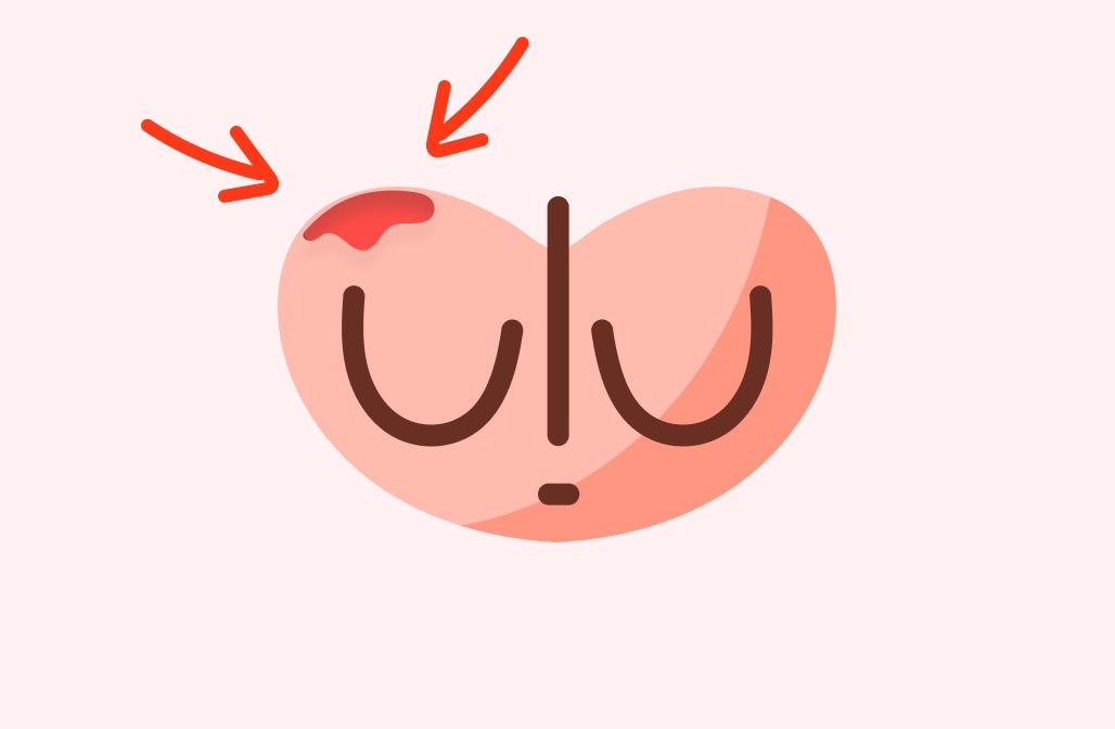
  • Ulceration: The skin breaks down—painful, often bleeding, and at risk of infection, notably in areas like the lip or diaper region.
  • Bleeding: Usually modest, but sometimes more, if the lesion is bumped or in a vulnerable spot.
  • Infection: Secondary to breakdown, requiring careful local care and sometimes antibiotics.
  • Threats to function: Hemangiomas near the eye—risking vision; those near the mouth—feeding issues. A strike across the airway? Breathlessness or noisy breathing.
  • Liver involvement: Extensive skin hemangiomas can sometimes signal the presence of hemangiomas in the liver, raising the rare risk of hormonal disturbances or heart strain.
  • Cosmetics and psychology: Scarring, discolouration, or persistent marks—especially on the face—can touch self-image, later requiring support and sometimes minor cosmetic corrections.

Prognosis and Outlook: The Future for Children with Infant Hemangioma

Good news: time is a friend more often than a foe. In the great majority, infant hemangioma shrinks away—sometimes within a few years, often by age ten. Residual changes? Rarely concerning—slight pallor, a gentle dip, a map of fine vessels. What is important: careful observation and not missing the few cases where rapid change, ulceration, or functional trouble emerge. If the appearance troubles you or your child, small interventions—sometimes dermatological, sometimes emotional support—are available.

Treatments and Management: From Watchful Waiting to Active Therapy

Most infant hemangioma needs no interference—just reassurance and regular check-ups:

  • Observation: Gentle watchfulness, especially with small, uncomplicated spots.
  • Pharmacological options:
  • Oral beta-blockers (propranolol): The mainstay for problematic, rapidly growing, or function-threatening marks. Early initiation is key, with medical monitoring.
  • Topical beta-blockers (timolol): For petite, superficial hemangiomas, these eye drop-like gels offer a gentle, targeted approach.
  • Corticosteroids: Used infrequently now, yet they have a place if beta-blockers are not suitable.
  • Other therapies: For stubborn cases, medicines like vincristine or sirolimus might be tried under specialist guidance.
  • Topical creams: Occasionally effective for minor lesions.
  • Laser therapy: Pulsed dye lasers target superficial or post-healing redness, especially on the face.
  • Surgery: Rare, but reserved for persistent, function-impairing, or ulcerated growths that defy medicine.
  • Supportive care: Skincare, wound healing, pain management, plus emotional reassurance for families faced with persistent or visible lesions.

Special Considerations: Unique Cases in Infant Hemangioma

Special care for special situations. Premature babies and those with low birth weight sit at higher risk, deserving closer checks. Marks suggestive of PHACE syndrome or those near vital organs demand a broader clinical team—paediatrician, dermatologist, sometimes neurologist or cardiologist.

Lesions with a “beard” pattern (chin, neck, lower face) ring warning bells for airway involvement—ENT consultation is wise. Near the eyes? An ophthalmologist’s evaluation is essential, as visual development is sensitive even to minor encroachments.

Multiple marks signal internal checks: liver scans, heart monitoring, and holistic consideration of the child’s growth, feeding, and development.

Current Recommendations and Evolving Science

Contemporary management champions an individualised, risk-based approach:

  • Low-risk: Routine review every 4–6 weeks in infancy.
  • Complicated or risky locations: Early treatment, especially with beta-blockers, after cardiovascular assessment.
  • Adjunctive therapies: Laser or surgery, only as needed, tailored to the lesion’s impact on appearance or function.
  • Scoring and monitoring: Use of clinical scoring systems and photo documentation to track progress.
  • Family education: Helping parents recognise warning signs and understand medicines’ effects.
  • Interdisciplinary collaboration: From paediatrician to dermatologist, and when warranted, other specialists join for best outcomes.

Research is advancing: New therapies, alternative beta-blockers, targeted interventions, and the search for reliable biomarkers like VEGF are reshaping future care. Psychological research also reminds us—timely intervention can secure not just skin health, but self-confidence and emotional well-being.

Daily Management and Prevention Tips

No manual exists for preventing infant hemangioma—they are not the result of anything done during pregnancy or after birth. Preventing trauma is practical: keep the area clean, use gentle moisturisers, regularly trim fingernails to prevent scratching, and avoid irritation. Should signs of infection (redness, oozing, pain) or sudden changes develop, medical advice is recommended immediately.

Gentle daily routines—cleansing, moisturising, and attentive monitoring—are the most effective “treatment” for uncomplicated marks.

Supporting Your Child and Family Well-Being

Beyond the medical, living with infant hemangioma weaves a tapestry of emotions. Support is multifaceted:

  • Keep the affected area clean and soft.
  • Monitor for change: rapid growth, breakdown, or discharge.
  • Adhere to any prescribed treatments, and never skip appointments.
  • Arm yourself with simple, reassuring responses for curious questions from friends or relatives.
  • Engage with parent networks and support groups online or locally—shared experience provides comfort.
  • Honest, age-appropriate explanations at home foster acceptance as your child grows.
  • Work with teachers so schooldays are as supportive as home.
  • Professional counselling is available for children or families who find the psychological impact lingers.

Trust in collaboration—medical, familial, and social—lays the foundation for a thriving, confident future.

Key Takeaways

  • Infant hemangiomas are widely seen, generally benign, and most fade away naturally, though a small percentage need treatment if they affect health or essential functions.
  • Prompt recognition, ongoing observation, and when needed, early therapy help avoid complications and support healthy development.
  • Multiple treatment pathways exist; from “wait and watch” to medications, laser, or surgery, choices adapt to each unique case.
  • Comprehensive family understanding and leveraging expert resources enhances both medical and emotional wellbeing.
  • Consult your healthcare provider promptly for any skin changes in your child that worry you; and for guidance, download the Heloa app for free questionnaires and tailored health guidance.

Questions Parents Ask

Can infant hemangiomas appear after birth or do they always show at birth?

Many parents feel baffled—a mark that wasn’t there at birth, but suddenly pops up in those first weeks? Entirely normal. Infant hemangioma typically develops postnatally, growing rapidly soon after birth rather than being present from day one. No cause for alarm—delayed appearance is common and not a signal of something being wrong.

Are infant hemangiomas contagious or caused by infections?

Unease about spreading to siblings or being linked to infection is understandable. Rest easy—infant hemangioma is not contagious. Neither viruses nor bacteria play any role, nor does hygiene or anything parents did during pregnancy or after. It is a matter of unusual blood vessel growth, not infection.

Will my child have long-term problems if they had a hemangioma as an infant?

For most children, the chapter closes peacefully. Once an infant hemangioma has faded, the skin may retain a tiny difference in colour or texture—a pale area, a gentle dip, sometimes a few fine vessels. These marks are almost always minor, rarely causing trouble later. Rarely, large or deeply located hemangiomas might leave more obvious changes, but health and growth remain unaffected. For any worry about lasting impact, discussing options with your paediatrician brings reassurance and, if needed, tailored suggestions.

Further reading:

Similar Posts

The moment you spot a vivid red spot blooming on your baby’s skin, a thousand worries can rush in at once. Why is this appearing? Will it harm my child? Should I act immediately or just wait and see? The appearance of an infant hemangioma, also known as a “strawberry mark,” certainly raises questions for many parents—questions that deserve clear, supportive answers, not just medical jargon.
Understanding what these vascular tumors are, how they develop, and what modern science reveals about their causes can make a world of difference in how you feel about your child’s skin and health. Dive into the details: what triggers an infant hemangioma, its typical course, when care is essential, and which reassuring advances shape how doctors approach treatment and support. Each section below will shine a light on different facets of this common childhood condition, blending up-to-date medical knowledge with practical advice to help you navigate each stage with confidence.

What Is an Infant Hemangioma? Signs and the Science Behind the Red Mark

Spotting an infant hemangioma—that unmistakable bright red, raised “strawberry patch”—can spark concern, curiosity, or both. At its core, an infant hemangioma is a benign vascular tumor made up of an increased number of tiny blood vessels, or capillaries, usually forming close to the skin’s surface. Unlike other birthmarks or persistent vascular anomalies like port wine stains, infant hemangiomas follow a dynamic life cycle. Typically, they erupt within the first months, race through a rapid growth phase (the “proliferative phase”), stabilize, and then—almost like a time-lapse flower closing—gradually fade during early childhood. Many parents find it startling at first, only to watch in wonder as the color and bumpiness diminish over time.

But why does this happen? Science points to special markers in these growths—like GLUT-1, a protein shared with placental tissue—that distinguish them from other skin lesions. This unique signature even assists doctors in making a precise diagnosis, especially when other birthmarks appear similar.

What makes infant hemangioma especially distinctive is its pattern: explosive early growth followed by slow, spontaneous shrinking (“involution”). For most, this cycle ends with little more than a faint trace on the skin by primary school age. Yet, the psychological comfort in understanding their typical arc shouldn’t overshadow rare cases where size, position, or complications require closer attention.

What Triggers an Infant Hemangioma? Genetics, Environment, and Risk Factors

If you’re wondering what sets the stage for an infant hemangioma, you’re not alone. Research is ongoing, but certain risk factors are now clear.
First, the genetic puzzle: while most cases remain random, rarely hemangiomas can appear in families, suggesting a potential hereditary element. Significantly, girls are up to five times more likely to develop these marks, though the reason for this sex difference is still being unraveled.

Let’s focus on known risk boosters:

  • Prematurity and low birth weight: The smaller and more premature a newborn, the higher the odds.
  • Maternal factors: Advanced age, multiple pregnancies (twins or more), or pregnancy complications like chorionic villus sampling subtly elevate risk.
  • Perinatal hypoxia: Reduced oxygen levels around birth spur the release of growth signals—angiogenic factors like VEGF—leading to excessive blood vessel formation.

Occasionally, infant hemangioma appears as part of more complex syndromes such as PHACE (affecting the brain, heart, and eyes) or LUMBAR (impacting urinary, skeletal, or nervous systems). In these settings, symptoms stretch far beyond the skin, emphasizing the need for a thorough, multidisciplinary evaluation.

How Does an Infant Hemangioma Evolve? Phases, Markers, and Medical Insight

Every infant hemangioma embarks on its own timeline, but the three core phases emerge almost universally:

  1. Proliferative phase (birth to 6–12 months): Supercharged growth as endothelial cells (the architects of blood vessels) multiply rapidly.
  2. Plateau phase: Growth rate plateaus, fewer new vessels form, and the lesion often remains unchanged for months.
  3. Involutional phase: The once-prominent mark softens, shrinks, and the bright dye fades—sometimes leaving behind mild skin changes, such as faint blood vessels or fatty tissue.

Molecular markers—like CD31 and particularly GLUT-1—help pathologists pinpoint the true origin of the growth, while high levels of VEGF highlight the underlying biology: the body’s delicate machinery for blood vessel formation going temporarily into overdrive.

How to Spot an Infant Hemangioma: Appearance, Patterns, and Locations

Noticing a cherry-red bump, bluish swelling, or a blend of hues on your child’s skin? The contrasts hint at depth:

  • Superficial hemangiomas: Sitting atop the skin, often lumpy, bright red, and well-outlined—the classic “strawberry” appearance.
  • Deep hemangiomas: Buried below the surface, they often look bluish or purplish, slightly squishy, and harder to spot when small.
  • Mixed hemangiomas: Combining both visible and underlying features.

The head, neck, eyelids, and hands score as the most common sites, but an infant hemangioma can develop anywhere—even occasionally on internal organs like the liver. Parents frequently report first spotting a faint patch of pink or white that balloons in size during the first six months.

Patterns matter too:

  • Focal: One single, defined lesion.
  • Segmental: A wider, band-like array along an anatomical region.
  • Multifocal: Multiple spots scattered across the skin.

Diagnosing Infant Hemangiomas: Clinical Acumen and When to Worry

For most children, a brief exam by a healthcare professional clinches the diagnosis of infant hemangioma. Timing, color, and shape provide plenty of clues. But what if the lesion is atypical, buried, or alarmingly close to the eye or mouth?

Here, imaging steps in:

  • Ultrasound with Doppler: Fast, painless, and precise for assessing blood flow.
  • MRI: Chosen for large, deep, or potentially syndromic marks.
  • CT scans: Rarely required, given radiation risks.

A biopsy—where a small sample is examined under the microscope—serves as a last resort, mainly to confirm ambiguous cases. Identifying that telltale GLUT-1 marker can serve as a medical fingerprint, cementing the diagnosis.

If large, fast-growing, or located in tricky places, further screening assesses the potential impact on vision, breathing, or internal organs, and guides the next steps.

Complications of Infant Hemangioma: Staying Ahead of Possible Problems

The reassuring news: most infant hemangiomas mean no harm and simply fade with time. Still, several complications can signal the need for hands-on management:

  • Ulceration: Pain, bleeding, and infection flare up when the skin breaks, especially on lips, diaper areas, or zones that rub against clothing.
  • Significant bleeding: Uncommon, but should never be ignored—bleeding is usually minor, yet trauma to these vascular marks requires prompt attention.
  • Infection: More frequent after ulceration, needing regular gentle hygiene and perhaps medical care.
  • Functional risks: If an infant hemangioma crowds the eye, ear, mouth, or airway, normal functions (vision, feeding, breathing) may be at risk.
  • Internal involvement: Multiple hemangiomas on the skin may signify hepatic hemangiomatosis—an overgrowth inside the liver that, in rare cases, affects hormones or heart function.
  • Cardiac load: Exceptionally large or abundant hemangiomas may place undue stress on the infant’s developing heart.
  • Aesthetic and psychological impact: Most fade, but the emotional toll—from teasing to self-consciousness—can linger, underlining the importance of early, open communication.

Prognosis: What Lies Ahead for Children with Infant Hemangiomas?

Let’s step back and look at prognosis. Overwhelmingly, infant hemangiomas shrink on their own, usually resolving by the time your child starts primary school. While port wine stains linger lifelong, infant hemangiomas usually fade away, demanding little intervention except monitoring. Watch for new symptoms, pigment changes, bleeding, or swelling and consult your provider promptly for peace of mind.

Treatment Approaches: From Watchful Waiting to Modern Medical Therapy

How to handle an infant hemangioma often depends on its size, speed of growth, and location. Rest assured, the approach is never a “one-size-fits-all.”

First-line care?
Often, simple observation. Most hemangiomas do not require immediate intervention. But some scenarios call for more than watchful waiting:

  • Oral beta-blockers (propranolol): The current favorite for problematic hemangiomas, well-tolerated and typically effective when given early under medical supervision.
  • Topical beta-blockers (timolol): Targeted for smaller, superficial marks, with promising results and minimal systemic effects.
  • Steroids (corticosteroids): Less in favor these days, but still valuable for select patients when beta-blockers are not advised.
  • Other medications: For resistant or especially severe cases—drugs like vincristine, sirolimus, and interferon might be introduced under expert care.
  • Laser therapy: The pulsed dye laser helps refine port wine stains or treat ulcerated, persistent, and sometimes disfiguring marks.
  • Surgical removal: Rarely required and reserved for complex, unresponsive, or functionally threatening lesions.
  • Supportive care: Never underestimate the benefit of meticulous skin care, wound management, pain relief, and robust family reassurance for overall quality of life.

Special Considerations: When Infant Hemangioma Signals Need for Extra Care

Some babies and scenarios deserve extra vigilance:

  • Premature and low-birth-weight infants: Rapid or atypical growth is more likely, and monitoring is even more essential.
  • Syndromic forms: In PHACE or LUMBAR, wider evaluation and multi-specialist follow-up are required.
  • Periocular and airway hemangiomas: Located near eyes or within the airway, these can challenge vision or breathing, making swift assessment by an ophthalmologist or ENT essential.
  • Multiple hemangiomas: Presence on the skin often prompts abdominal ultrasound to detect any internal ones, especially in the liver.

Sensitive sites—particularly the face or neck—carry weight for self-confidence and social inclusion as children grow, reinforcing the need for psychological support alongside medical care.

Day-to-Day Care Tips and Prevention: Keeping Your Child Comfortable

Predicting or preventing infant hemangioma is, unfortunately, not possible. Their emergence is not linked to parental actions before or after birth. Still, daily care makes a difference:

  • Maintain gentle hygiene: Clean, well-moisturized skin is less likely to ulcerate or become infected.
  • Prevent scratching: Keep nails trimmed and soft mittens handy for little hands prone to exploring.
  • Monitor for changes: Watch for any rapid size increase, color shifts, or pain—each cue to consult your healthcare provider.

Small adjustments in your routine can help avoid irritation, minimize discomfort, and give you a sense of agency in your child’s daily comfort.

Family Well-Being and Emotional Support: The Human Side of Infant Hemangioma

Parenthood often brings a storm of emotions, especially when visible changes appear on your child’s skin. Accepting and understanding an infant hemangioma means balancing clinical facts with lived experience:

  • Be proactive: Stick to recommended check-ups and attend to medication schedules as advised by your care team.
  • Prepare positive explanations: Reassure your child (and others) with simple, honest answers about the visible mark.
  • Connect to support: Parent groups, informative websites, and counseling can transform feelings of uncertainty into empowerment.
  • Foster open conversation at home and school: Honest dialogue lays the foundation for resilience and confidence, even in the face of curiosity or ignorance from others.
  • Seek psychological support if needed: Professional insight is invaluable when appearance-related issues affect you or your child’s self-image.

Open communication, a strong support network, and daily routines foster an environment where your child can thrive, mark or no mark.

Key Takeaways

  • Infant hemangioma is a common benign vascular growth that usually follows a predictable arc: rapid growth, plateau, and slow regression.
  • Most cases need only gentle observation, while some require prompt intervention, especially when vital functions are at risk.
  • Treatments are tailored: from doing nothing but watching, to using oral or topical medications, laser therapy, or—on rare occasions—surgery.
  • Advances in molecular understanding and treatment choices empower parents and physicians alike to provide effective, compassionate care.
  • Emotional support for both child and family is as important as the medical approach.
  • For any new or evolving spot on your child’s skin, consult a qualified professional for assessment and reassurance.
  • To access ongoing expert advice and free pediatric health questionnaires, download the Heloa app—a valuable ally on your parenting journey.

Questions Parents Ask

Can infant hemangiomas appear after birth or do they always show at birth?

It’s common for parents to notice that hemangiomas can develop after birth rather than being present immediately. Most infant hemangiomas typically become visible within the first few weeks of life, rather than being there at delivery. If a mark or bump appears a little later, pas d’inquiétude — this timing is entirely normal and doesn’t indicate anything unusual about your child’s health.

Are infant hemangiomas contagious or caused by infections?

Rassurez-vous, infant hemangiomas are not contagious and cannot be spread from one child to another. They aren’t the result of an infection, nor do they reflect anything a parent did or didn’t do during pregnancy or afterward. Hemangiomas are due to an overgrowth of blood vessels and have no link to hygiene, viruses, or bacteria.

Will my child have long-term problems if they had a hemangioma as an infant?

Most children with infant hemangiomas grow up without any lasting health concerns. Once the hemangioma has faded, the skin may have a slight difference in color or texture, but this is rarely bothersome. In a few cases, especially if a hemangioma was very large or located in a sensitive area, small marks or mild skin changes may persist. However, these do not usually impact a child’s health or development. If you have any worries about the long-term appearance or well-being of your child, n’hésitez pas à en parler avec un professionnel de santé.

Further reading:

Similar Posts