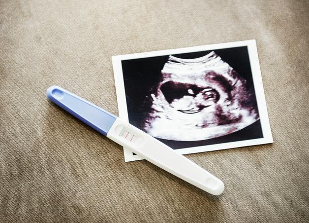
Few moments are as stirring for expecting parents as watching those hazy, flickering life signs on the ultrasound monitor for the very first time. Curiosity swirls alongside the thrum of anticipation—Will there be a heartbeat? Is everything positioned well inside the uterus? Am I really at the right number of weeks? The first trimester ultrasound stands as both a guardian and a witness: it confirms pregnancy location, listens for cardiac activity, estimates due dates with unmatched accuracy, and quietly searches for hints of twins or the rare marker that might need extra attention down the line. For families navigating early pregnancy uncertainties—anxious over dreamlike symptoms, bracing for relief—this early window offers concrete, visual reassurance. You might wonder, “What exactly will they see? Will answers be immediate? How soon will I know everything is on track?” Each of these puzzles finds its place as the ultrasound probe traces gentle circles and captures those first grainy glimpses of a new life. Let’s explore what a first trimester ultrasound reveals, how the experience feels, what results you might encounter, and how each detail shapes your path forward.
Why the first trimester ultrasound matters for parents
Regardless of whether this is your first or fifth pregnancy, the need for clarity echoes in every waiting room. The first trimester ultrasound delivers on this need for certainty, providing evidence that the pregnancy is developing as expected (or, gently, that a change of expectations is needed). Key details come alive during this scan:
- Confirming viability—that is, you’ll actually see if the baby’s heart is beating, and whether the pregnancy is safely settled inside the uterus (not outside, where complications are more likely).
- Accurate dating—the crown–rump length (CRL) measured on ultrasound during this stage beats any calendar calculation by several days, resetting your estimated due date if needed.
- Screening early for potential concerns—features like nuchal translucency (the small pocket of fluid behind baby’s neck), when measured at the perfect time, offer a non-invasive way to check for some genetic conditions without any discomfort to your baby.
- Guiding next steps—from when to repeat scans and whether extra tests are helpful, to pinpointing when to get in touch for pain or bleeding, this scan is a starting point for careful, personalised care.
Purpose and timing: first glimpses, first answers
Early viability and dating (6–10 weeks)
Here, the first trimester ultrasound often uses a transvaginal probe—think of it as a slim, covered wand, providing crisp views when everything is still tiny. In just a few minutes, the sonographer checks for a gestational sac (that first bubble of pregnancy), a round yolk sac (the embryo’s first source of nutrients), a hint of the fetal pole, and that all-important flicker of cardiac motion. When measured between six and ten weeks, the CRL gives a highly precise estimate for your due date—better than remembering last period dates that often blur with time.
Nuchal translucency window (11–13 weeks + 6 days)
Most clinics suggest the main first trimester ultrasound within this window. The baby has now grown enough for key measurements: crown–rump length, heartbeat, and nuchal translucency if you opt for early genetic screening. For families using IVF or similar techniques, an extra scan a few weeks after embryo transfer often checks for pregnancy location and viability, and a later first trimester ultrasound focuses again on the NT and growth markers.


Common reasons to schedule a first trimester ultrasound
- Unexpected or heavy bleeding, or any pelvic pain
- Confirming this is a viable pregnancy inside the uterus
- Estimating how many weeks along you are (especially if periods were irregular)
- Checking if you’re expecting twins (or more), and if so, what kind
- Measuring NT as part of first trimester screening
- Following up after uncertain or abnormal initial blood tests
What clinicians check and record
Your ultrasound report won’t just say “all is fine.” Each detail is considered:
- Gestational sac: location, size (measured as mean sac diameter or MSD), and presence
- Yolk sac: shape and size, looking for the healthy, rounded form
- Fetal pole and heartbeat: counted and documented, with fetal heart rate (FHR) typically 90–110 bpm at the earliest, rising to 120–170 bpm by the end of the trimester
- CRL for dating—the gold-standard measurement for this stage
- Uterus and ovaries: checking for fibroids, cysts, or anything unusual like a subchorionic hematoma (a collection of blood that is common but usually resolves)
- Special attention if concerns arise, such as possible molar pregnancy (unusual growth pattern inside the uterus) or findings that might suggest an ectopic pregnancy
Ultrasound types: transvaginal and transabdominal
Transvaginal ultrasound: precision’s early ally
Might sound a little daunting at first, but for ultra-early pregnancy, nothing beats the clarity of a transvaginal scan. A covered probe is gently inserted, and after a few seconds of mild pressure or cramping, high-quality images reveal the earliest details. Privacy is respected, you may bring a support person, and the process is usually quick—less than ten minutes for most.
Transabdominal ultrasound: the classic view
As pregnancy progresses (even after just a few weeks), a transabdominal first trimester ultrasound—applied via your tummy with some cool gel—often provides enough information. A not-too-full, not-too-empty bladder is the best friend of this technique, as it helps make the uterus easier to visualise. Limitations exist if your pregnancy is still very early or the position is tricky, but for most, this is a comfortable, routine part of pregnancy care.
What does the appointment feel like?
Expect about 20–30 minutes for the scan itself. You’ll see the screen, often as the sonographer explains what appears—“Here’s the gestational sac… and now the heartbeat!” There’s always space for questions, and if a nuchal translucency measurement is needed, you may be asked to move or cough, encouraging baby into the best position.
Early findings: what parents want to know
CRL and dating accuracy
No guessing here. The neutral-position CRL on first trimester ultrasound is the single most accurate way to settle the due date—a small difference from your period dates is common and usually nothing to worry about.
Gestational sac, MSD, and yolk sac
Before baby is visible, the gestational sac and its MSD (average size, measured in millimeters) offer clues as to how old the pregnancy really is. A yolk sac should look round and proportional; an odd shape or very large yolk sac has a stronger chance of signalling a possible miscarriage, so close follow-up is considered.
Cardiac activity and normal heart rates
Typically, a heartbeat appears on ultrasound as early as 5.5 weeks (using transvaginal method), rising from around 90–110 bpm at six to seven weeks, and stabilising near 120–170 bpm later. No heartbeat when one would be expected? Waiting seven to fourteen days and repeating both ultrasound and blood tests often clarifies things.
Nuchal translucency (NT): what does it mean?
The nuchal translucency is a small clear area at the back of baby’s neck. Measured between 11 and 13+6 weeks, an increased thickness sometimes suggests a higher chance for certain chromosomal conditions (such as Down syndrome), but it is not diagnostic on its own. This measurement is done by trained staff under strict protocols—you might be asked to wait, move, or try a gentle cough to help baby get into position.
Early anatomy
Some major structural changes can be spotted even this early. Absent cranial vault (acrania), large abdominal wall openings, or cystic hygroma (a specific swelling in baby’s neck)—these rare findings prompt a quick referral to specialists, and plenty of support is provided.
First trimester screening: tests and techniques
Combined first trimester screening
A first trimester ultrasound with NT measurement, combined with blood markers (PAPP-A and free beta-hCG) and maternal age, predicts a numerical risk score for chromosomal conditions (such as trisomy 21, 18, and 13). It’s a probability—not a “yes/no” answer. Both before and after, time is given to explain what the numbers mean, allaying fears and offering a clear route through the next choices.
Non-invasive prenatal testing (NIPT)
Parents seeking more accuracy—often for peace of mind—can opt for NIPT. This test captures fragments of placental DNA floating in the mother’s blood (from around 9–10 weeks), offering a high degree of confidence for detecting common chromosomal anomalies, including trisomy 21. For some, NIPT is an added or standalone screening; for others, it may be reserved for higher risk or after an initial screen.
Follow-up after a high-risk screen
When a screen result points to higher risk, options include another round of risk assessment (with NIPT) or diagnostic confirmation with chorionic villus sampling (CVS) late in the first trimester, or amniocentesis in the second. Dedicated professionals explain the process, risks, and what each test actually checks—making it easier to understand where you stand and what happens next.
What the first trimester ultrasound can—and cannot—reveal
- What it can show: gestational sac and yolk sac, embryo, fetal heartbeat, highly accurate CRL, NT thickness, and a handful of larger structural changes
- What it cannot show: fine details, which become clearer only in later scans—technical factors like baby’s position and even maternal build can influence what’s visible
Even a thicker-than-average NT is not a diagnosis: it prompts only deeper discussion, possible further blood tests, or perhaps targeted procedures based on your unique picture.
When things don’t go as planned: early complications
- Anembryonic gestation or missed miscarriage: sometimes the gestational sac grows, but remains empty, or the baby stops growing after previous heart activity. Borderline cases mean waiting and repeating tests before acting, often seven to fourteen days apart.
- Ectopic pregnancy or pregnancy of unknown location: if a first trimester ultrasound finds an empty uterus but an abnormal mass or fluid in the pelvis, swift action and close monitoring follow. Heavy pain or bleeding? Immediate medical care is warranted.
- Molar pregnancy: a very specific, unusual pattern in the uterus alongside markedly increased hCG points here, and management moves forward with coordination.
- Multiple pregnancies and chorionicity: early scans spot whether twins share the same placenta or not, shaping follow-up. Dichorionic (two placentas) is less risky than monochorionic (one shared placenta)—a “lambda sign” suggests the former, a “T-sign” the latter.
- Subchorionic hematoma or adnexal cysts: a small blood collection by the sac is fairly common and often harmless. Large hematomas need closer observation.
Safety, preparation, and your experience
Safety
First trimester ultrasound utilises sound waves, not X-rays. No evidence exists that it harms baby or parent when operated at medical standards and with proper duration and settings. Early Doppler use is limited to particular indications.
Preparation tips
- Bladder matters: moderately full makes transabdominal scans clearer, an empty bladder is preferred for transvaginal.
- Dress in comfortable clothes, allow easy abdominal access.
- Carry any earlier test reports or relevant paperwork—especially if referred from a fertility clinic or have recent blood test results.
- Jot down questions or worries; every inquiry is valid and deserves a careful answer.
The emotional spectrum is part of the experience
Everyone processes these moments differently. Some laugh, a few quietly cry, others simply grip their partner’s hand until results are read. Your thoughts, doubts, and hopes all have space here. If having a chaperone or a trusted support person soothes nerves, you are free to ask.
When the scan is unclear—next steps
If visualization is poor—perhaps due to very early timing, a tilted uterus, body shape, or scar tissue—a plan emerges: repeat imaging after seven to fourteen days (often paired with blood tests), plus clear advice on what symptoms should trigger prompt contact.
Practical checklists for parents
Before the scan
- Carry all previous test results and fertility clinic paperwork.
- Follow bladder instructions provided by your clinic.
- Wear clothing suitable for abdominal access.
- Make a list of queries—on dating, screening, next appointments, and emergency contacts (especially for pain or heavy bleeding).
- Mental preparation: nerves are normal. A support person or chaperone can make a big difference.
After the scan
- Clarify the significance of every measurement.
- Ask about follow-up appointments or further imaging—get clarity on when and how results reach you.
- Know who to call—bleeding, fever, or severe pain all require clear instructions for emergencies.
- If screening is performed, note when to expect those results and what kind of support or counseling is offered if the risk requires attention.
Understanding your ultrasound report
- Abbreviations you’ll encounter: CRL (crown–rump length), GA (gestational age), EDD (estimated due date), FHR (fetal heart rate), GS (gestational sac), MSD (mean sac diameter), YS (yolk sac), NT (nuchal translucency)
- Remember, risk scores from screening provide probabilities, not final answers. High risk leads to offers of further testing—including NIPT (accurate, noninvasive), CVS, or amniocentesis (both diagnostic but carrying small risks).
Logistics, costs, and getting your report
A first trimester ultrasound can be done in a variety of centers—maternity clinics, radiology departments, or specialised fetal medicine units. Experienced sonographers carry out the scan, and results are shared by the healthcare team directly or via digital patient portals. NT measurements demand special training for accuracy. Prices vary widely; ask for the expected out-of-pocket costs and whether you’ll receive a printout or digital copy. Clinical images always trump keepsake pictures; for personal mementos, check if your provider offers a print.
Key Takeaways
- First trimester ultrasound (typically 6–13+6 weeks) is the key to confirming healthy pregnancy, exact dating, early detection of twins, and safe first trimester screening.
- Most have the main scan between 11 and 13+6 weeks, but earlier ultrasounds support those with pain, bleeding, prior losses, or IVF treatments.
- A “normal” scan gives real comfort, knowing many details become visible only later in pregnancy.
- Sound waves, not radiation: first trimester ultrasound is trusted and safe when used as recommended by qualified professionals.
- Prepare well—bring earlier notes, manage your bladder, have those questions ready—and expect honest, respectful communication.
- Uncertain results or incidental findings aren’t verdicts; they’re guideposts for next steps and follow-up.
- Professional guidance is readily available. And for ongoing support, insights, and even tailored child health questionnaires, download the Heloa app for expert-backed resources and a personalised journey.
Questions Parents Ask
Can my baby’s sex be determined in the first trimester?
Everyone wants to know, right? In routine first trimester ultrasound, determining fetal sex reliably is not possible. Sometimes, people talk about “nub theory” (the angle in early scans)—but scientifically, it’s not conclusive until the anatomy scan, usually around 18–20 weeks. However, if knowing early is extremely important to you, non-invasive prenatal testing (NIPT), available from 9–10 weeks onwards, can typically identify the baby’s sex using DNA in the mother’s blood. Discuss this with your doctor to decide the best approach for your family.
When and how will I get my scan and screening results?
Usually, some of the findings like location, heartbeat, and dating are shared in real-time during the scan. A formal report along with the images might reach you via patient portal or email within a few days. For blood-based screening (including NT and first trimester combined screening), results are typically available a few days after your sample is processed. NIPT reports may take between 7–14 days. Diagnostic test results—for those who go ahead with amniocentesis or CVS—might arrive within 1–2 weeks. If any risk is noted, clear counseling and follow-up appointments are set up, so you are supported at every step.

Further reading :