
Pregnancy and early parenting come with a very particular kind of curiosity. You can feel your baby changing, yet you cannot see it day by day. So when a scan report, an app, or a well-meaning relative mentions baby size, the mind immediately starts doing maths: “Is it fine?”, “Is it small?”, “Is it too big?”
Here’s the steadying truth: baby size is usually an estimate. Useful, yes. Perfect, no. In India, as elsewhere, clinicians rely on the story across time: growth trend, placenta support, amniotic fluid, and your health.
Baby size in pregnancy: what parents can expect
What “baby size” means during pregnancy (curiosity vs clinical meaning)
During pregnancy, baby size commonly means an ultrasound-based estimate of fetal growth. In medical language you will hear estimated fetal weight (EFW), calculated using measurements such as head size, tummy size, and thigh bone length.
Curiosity is natural. But clinically, baby size is not a direct weighment like on a scale. It is a best-possible approximation, and what matters most is whether your baby is growing steadily.

Baby size vs gestational age: related, but not the same
Gestational age is the pregnancy timeline (often counted from the last menstrual period, then refined by an early ultrasound). Baby size usually rises with gestational age, so growth charts compare your baby with others of the same age.
Still, the two are not interchangeable. A baby can be smaller or bigger than “average” and be healthy. Your clinician reads baby size along with:
- pregnancy dating accuracy
- growth pattern over time
- placenta function
- amniotic fluid
- your medical history (thyroid, blood pressure, diabetes, etc.)
Why baby size estimates can change from week to week
Seeing a percentile shift, especially late in pregnancy, can feel unsettling. Common reasons are often technical:
- ultrasound has built-in variability, a few millimetres can change EFW and percentile
- different weight estimation formulas can give different EFW
- baby’s position and movement change the view
- small dating differences can shift percentiles
That is why doctors focus on growth velocity across repeat scans, rather than treating one report as a final judgement.
How clinicians measure baby size during pregnancy
First trimester: crown-rump length (CRL)
In early pregnancy (roughly 6-13 weeks), crown-rump length (CRL) is the key baby size measurement. CRL is also the most accurate single ultrasound measure for dating. Good dating becomes the reference point for later growth comparisons.
Second and third trimester: fetal biometry
From the second trimester onwards, clinicians use fetal biometry:
- BPD (biparietal diameter): head width
- HC (head circumference)
- AC (abdominal circumference)
- FL (femur length)
Each tells a slightly different story. AC often strongly influences calculated weight because it reflects soft tissue and liver size, which respond to nutrition and placental supply.
Estimated fetal weight (EFW): helpful, not exact
EFW is calculated by plugging HC/BPD, AC and FL into validated equations (often Hadlock-based). It helps screen for growth concerns, decide follow-up timing, and sometimes assist delivery planning.
But it is not an exact reading. In late pregnancy, the margin of error can widen, so the trend of baby size over time is more informative than a single EFW.
Percentiles and growth charts
Percentiles compare your baby with a reference group at the same gestational age:
- 50th percentile: roughly half of babies are smaller and half are larger
- many healthy babies lie between the 10th and 90th
A baby staying near the same percentile over time is often reassuring. Crossing several lines, or slowing growth velocity, may lead to closer monitoring.
Fundal height: a screening check
Fundal height is measured using a tape from the pubic bone to the top of the uterus (often after 20 weeks). A common thumb rule is that the measurement in centimetres roughly matches weeks of pregnancy in mid-to-late pregnancy (often within plus/minus 2 cm).
It can be influenced by fetal position, amniotic fluid, fibroids, multiples, placenta location, and body build. If it remains consistently high or low, ultrasound is usually used to check growth and fluid.
Baby size by week: milestones, charts, and calm interpretation
Week-by-week charts help you visualise the general pattern. They do not predict exact baby size on a particular day.
To keep charts helpful:
- treat numbers as ranges, not targets
- remember different sources use different charts and EFW formulas
- give priority to repeat clinical measurements over app updates
If something looks surprising, ask: was the estimate based on CRL or on HC/AC/FL? Which chart is used in your hospital? How does today compare to earlier scans?
First trimester: fast development, dating focus
Early weeks are about organ formation and rapid change. Clinically, the focus is usually accurate dating and confirming expected early development.
Second trimester: proportion and organ maturation
Second trimester growth brings more proportion. Many parents in India have the anomaly scan during this phase. Biometry becomes more meaningful, and movements feel stronger.
Third trimester: weight gain and fat stores
Third trimester is where weight gain accelerates as fat stores build and the lungs and brain continue maturing. Monitoring focuses on whether the baby is gaining appropriately and whether placental support remains adequate.
Fruit comparisons: enjoyable, not medical
Fruit comparisons are popular because they are easy to imagine and easy to share. They communicate one idea well: your baby is growing.
They do not capture what doctors track: growth velocity, placenta function, amniotic fluid, and sometimes Doppler blood flow.
So if a fruit comparison makes the baby size sound “too small”, take a breath. It is a rough visual, not a clinical conclusion.
What can influence baby size in the womb
Genetics and family traits
Genetics strongly shape growth potential. A petite couple may naturally have a smaller baby. A taller, broader-built couple may naturally have a bigger baby. Normal ranges also vary across populations, including within India.
Placenta function and oxygen/nutrient delivery
The placenta is the baby’s supply line. When placental function is reduced, growth can slow, sometimes leading to fetal growth restriction (FGR). If there is concern, clinicians may add Doppler studies and more frequent scans.
Nutrition and pregnancy weight gain
Adequate calories, protein, iron, folate, iodine and overall balanced nutrition support healthy growth. Very low maternal weight gain or malnutrition can be linked to smaller babies. Excessive weight gain, especially with diabetes, can be linked to larger babies.
If nausea, vomiting, heartburn, food aversions, or limited access to varied food is affecting intake, discuss it early with your doctor.
Lifestyle exposures
Smoking in pregnancy is associated with reduced fetal growth. Alcohol exposure can affect growth and development. If stopping feels difficult, ask for help.
Maternal health conditions
Some conditions often travel with particular baby size patterns:
- diabetes (including gestational diabetes) can be associated with larger babies
- hypertension and related disorders can increase the chance of growth issues in some pregnancies
Multiples (twins, triplets)
Twins and higher-order multiples often have lower individual weights compared with singletons at the same gestational age. Growth expectations and charts differ.
Normal variation between ultrasounds
Even in healthy pregnancies, two ultrasounds may not match perfectly. Fetal position, amniotic fluid, equipment settings, and millimetre-level differences can shift EFW. A modest change is common, a consistent trend is what carries meaning.
Percentiles explained: SGA, AGA, LGA
AGA: appropriate for gestational age
AGA usually refers to measurements roughly between the 10th and 90th percentile.
SGA vs FGR (IUGR)
SGA (small for gestational age) is typically below the 10th percentile. Some SGA babies are constitutionally small and well.
FGR (also called IUGR in older terms) suggests the baby may not be reaching their growth potential, often due to placental insufficiency. Doctors may look at growth velocity, Doppler blood flow, amniotic fluid, and the overall pattern.
LGA and macrosomia
LGA (large for gestational age) is often above the 90th percentile. Macrosomia usually refers to a high birthweight cut-off (often 4,000 g or 4,500 g depending on the definition).
Larger baby size may influence delivery planning because it can increase the risk of shoulder dystocia and birth trauma. Many LGA babies are healthy, the aim is planning.
When baby size needs a closer check
A growth ultrasound may be suggested when fundal height remains smaller or larger than expected, a previous scan suggests SGA or LGA, there are risk factors (diabetes, hypertension, multiples), or placental concerns.
A growth scan report often includes HC/BPD, AC, FL, EFW and percentiles, plus amniotic fluid assessment and placenta notes. Doppler studies may be added when needed.
Baby size after birth: growth beyond age labels
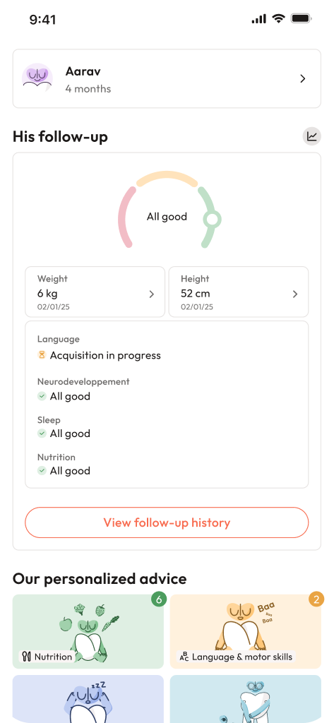
After delivery, baby size is never just one number. Paediatric follow-up uses:
- weight
- length (measured lying down until about 2 years)
- head circumference
These are plotted on WHO growth charts (separate for boys and girls). One practical combined view is weight-for-length, which reflects body build better than age alone.
Practical landmarks (wide normal ranges)
These ranges are broad, your child’s personal trajectory matters more:
- Newborn: around 50 cm, and commonly 3.3-3.5 kg on average (with wide normal range)
- 6 months: often roughly 63-71 cm
- 12 months: often roughly 71-80 cm
- 24 months: often roughly 83-93 cm
Clothing tip that saves time: if the label shows centimetres, follow that more than “months”, because body proportions vary a lot.
Measuring baby size at home (simple, consistent methods)
Weight
An infant scale is best. Measure at a similar time, with a similar diaper/clothing approach, and take 2-3 readings.
No infant scale? A bathroom scale can give a rough trend: weigh yourself holding the baby, subtract your weight alone.
Length (until about 2 years)
Measure lying down on a firm surface. Keep the head straight, align shoulders and hips, gently extend one leg without forcing, then measure head-to-heel. Measure twice and average.
Head circumference
Use a soft tape above the eyebrows and ears, around the largest part at the back of the head. Measure twice and average.
A practical rhythm:
- 0-6 months: about monthly
- 6-12 months: every 4-8 weeks
- 12-24 months: every 6-8 weeks
When to seek medical advice after birth
Speak to your paediatrician if you notice a clear break in growth trend, stagnation across several measurements, persistent rapid gain, or symptoms like poor feeding, unusual tiredness, vomiting, or diarrhoea.
If weight, length and head circumference seem mismatched, re-check measuring technique and discuss it. Context often explains the pattern.
Key takeaways
- baby size in pregnancy is an estimate, steady growth over time often matters more than one scan.
- Ultrasound uses CRL early, then HC/BPD, AC and FL to calculate EFW and percentiles.
- Genetics, placenta function, nutrition, exposures, medical conditions and multiples can all influence baby size.
- After birth, baby size is best understood using weight, length and head circumference together on growth charts.
- If anything feels unclear, your obstetrician or paediatrician can explain the trend and plan follow-up.
- You can download the Heloa app for personalised guidance and free child health questionnaires.

Further reading :