Edwards syndrome can arrive like a sudden pause in pregnancy—an unexpected word, a test result, an ultrasound comment that lingers in the mind. Parents often want two things at once: clear medical facts (without vague phrasing) and a sense of what decisions may look like, day by day, from pregnancy to the newborn period and beyond. The reality is that Edwards syndrome affects many body systems, yet every baby’s course remains individual. Screening results, confirmed diagnosis, the specific type of trisomy 18, and the care goals chosen with the medical team all shape what comes next.
Edwards syndrome: what it is, and why the name matters
Trisomy 18 in plain terms
Edwards syndrome is a genetic condition caused by an extra copy of chromosome 18 in some or all of a baby’s cells. Chromosomes are packages of DNA, they carry genes that guide development. When there is extra chromosome material, development can be altered in several organs at the same time.
There is no treatment that can “remove” the extra chromosome. Care is therefore supportive: treating symptoms when possible, preventing avoidable complications, and focusing on comfort and family goals.
“Edwards syndrome,” “trisomy 18,” “T18”: same diagnosis, different labels
You may hear several terms:


- Edwards syndrome
- Trisomy 18
- T18
Clinicians sometimes describe results with a karyotype, such as 47,XX,+18 or 47,XY,+18. That line simply means there are 47 chromosomes instead of 46, with an extra chromosome 18.
Screening versus diagnosis: two very different types of answers
Many parents are first told about Edwards syndrome through a screening result. That distinction matters.
- Screening estimates probability. Examples: ultrasound “markers,” blood tests, and cfDNA/NIPT.
- Diagnostic testing confirms or rules out the condition. Examples: CVS (chorionic villus sampling) or amniocentesis, followed by chromosome analysis.
A screening test can be highly sensitive, yet it still cannot give certainty on its own. A diagnostic test aims to provide certainty.
Types of trisomy 18: full, mosaic, and partial
Edwards syndrome does not always look the same because trisomy 18 can exist in different forms:
- Full (complete) trisomy 18: nearly all cells carry the extra chromosome 18. This is the most frequent form and, in general, the most severe.
- Mosaic trisomy 18: some cells have trisomy 18 and others do not. Signs may be milder, or simply more variable, depending on how many cells are affected and in which tissues.
- Partial (segmental) trisomy 18: only part of chromosome 18 is present in extra copies, sometimes due to an unbalanced translocation. Outcomes depend strongly on which segment is duplicated and how large it is.
You might wonder: “Does the type change what our baby will experience?” It can. Not always in a predictable way, but it often guides discussions about prognosis and care options.
Genetics, causes, and risk factors
Extra chromosome 18: aneuploidy and gene dosage
Trisomy 18 is an aneuploidy, meaning an atypical number of chromosomes. The additional chromosome changes gene dosage (extra copies of many genes), which can disrupt organ formation and growth.
How it happens: nondisjunction and early cell-division errors
Most cases of full Edwards syndrome happen due to nondisjunction—a separation error when an egg (more rarely a sperm) is formed. This is why risk increases with maternal age.
Mosaic trisomy 18 often results from an error after fertilization, during early cell divisions, creating a mix of typical cells and trisomic cells.
Partial trisomy 18 and translocations: when parental testing is discussed
Partial trisomy 18 can be linked to an unbalanced translocation (extra chromosome 18 material attached to another chromosome). Sometimes, a parent carries a balanced translocation—no health effects for them, but a higher chance of unbalanced chromosome patterns in a pregnancy.
When partial trisomy 18 is identified, clinicians may propose parental karyotypes to clarify recurrence risk.
Maternal age and “chance biology”
Maternal age is the best-established population-level risk factor. Risk rises more clearly from around age 35. Still, Edwards syndrome can occur at any age, often without any family history.
Recurrence risk: what families are commonly told
After a pregnancy with full trisomy 18, recurrence risk is often quoted around 1% (above the baseline age-related risk). If a translocation is involved, risk can be higher, which is why genetics input can be helpful.
How common is Edwards syndrome?
At conception, trisomy 18 is often estimated around 1 in 2,500 pregnancies. Live-birth prevalence is lower—commonly cited around 1 in 6,000 to 1 in 8,000—because many affected pregnancies end in miscarriage or stillbirth, and some families choose to end a pregnancy after confirmation, depending on personal circumstances and local laws.
Statistics can be useful for counseling, yet they cannot predict what will happen for one baby.
Prenatal screening and diagnostic tests
First-trimester and second-trimester screening
- First-trimester combined screening (around 10–14 weeks) uses nuchal translucency on ultrasound plus maternal serum markers (PAPP-A and free beta-hCG). It provides a probability estimate.
- Second-trimester quad screen is another probability estimate when first-trimester screening was not performed.
cfDNA/NIPT: strong screening, still not confirmation
cfDNA/NIPT can be done from about 10 weeks. It is very sensitive for trisomy 18, but false positives and false negatives can occur.
Why? A few examples:
- Low fetal fraction (not enough placental DNA in the blood sample)
- Confined placental mosaicism (placenta differs from fetus)
- Maternal factors (rarely)
A positive NIPT result for Edwards syndrome is usually followed by diagnostic testing.
Diagnostic testing: CVS and amniocentesis
- CVS is often performed around 10–13 weeks and samples placental tissue. It provides earlier information, though placental mosaicism can occasionally complicate interpretation.
- Amniocentesis is usually performed from around 15 weeks and tests fetal cells from amniotic fluid.
Both can confirm Edwards syndrome through chromosome analysis.
Karyotype, FISH, and chromosomal microarray (CMA)
- Karyotype confirms trisomy 18 and can indicate full, mosaic, or translocation-related patterns.
- FISH can provide a rapid preliminary answer, then is followed by full analysis.
- Chromosomal microarray (CMA) gives higher-resolution information and can be particularly informative for partial trisomy 18.
Diagnosis after birth
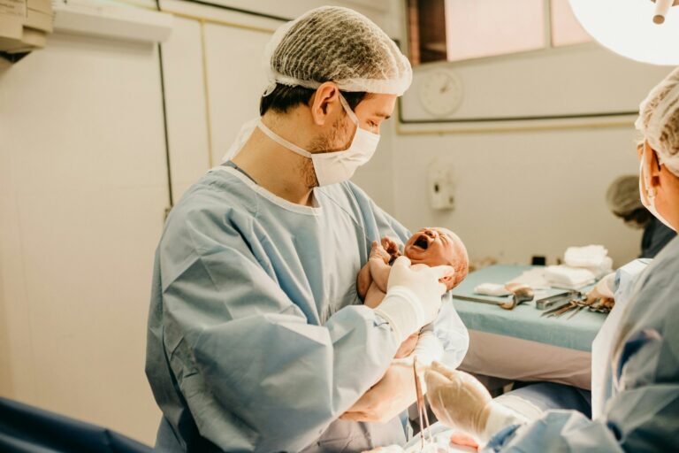
Sometimes Edwards syndrome is suspected after birth because of physical features and medical findings (for example, heart disease plus characteristic limb posture). Confirmation is made with chromosome testing from a blood sample.
Ultrasound findings that may suggest Edwards syndrome
No single sign is definitive
Ultrasound signs can increase suspicion, but an isolated finding rarely confirms Edwards syndrome. Clinicians look for a pattern.
Findings often discussed
Possible prenatal findings include:
- Intrauterine growth restriction (IUGR)
- Polyhydramnios (excess amniotic fluid)
- Congenital heart anomalies
- Clenched hands with overlapping fingers
- Omphalocele (abdominal wall defect) in some pregnancies
If such findings appear, a fetal echocardiogram (detailed heart ultrasound) may be proposed.
Signs and symptoms after birth
What parents may notice early
Many newborns with Edwards syndrome are small for gestational age and may have hypotonia (low muscle tone). Feeding can be difficult—poor suck, fatigue, and trouble coordinating suck-swallow-breathe.
Breathing can also be fragile. Sometimes it is subtle, sometimes it is immediate.
Heart defects
Congenital heart disease is common in Edwards syndrome. Examples include:
- VSD (ventricular septal defect)
- ASD (atrial septal defect)
- PDA (patent ductus arteriosus)
These can contribute to fast breathing, sweating or fatigue with feeds, poor weight gain, and oxygen needs.
Neurologic involvement and breathing instability
Neurologic differences are frequent. In full trisomy 18, development is typically profoundly affected.
Possible findings include:
- Structural brain differences (for example, agenesis of the corpus callosum)
- Apneas (pauses in breathing)
- Seizures in some babies
Limb and posture findings
A classic pattern is clenched fists with overlapping fingers. Clubfoot, “rocker-bottom” feet, stiffness, and contractures (limited joint movement) may be present.
Kidneys, digestion, and feeding safety
Kidney differences can occur, including hydronephrosis or horseshoe kidney, with possible urinary tract infections.
Digestive and feeding concerns may include:
- Gastroesophageal reflux
- Aspiration risk (milk entering the airway)
- Constipation
- Sometimes major malformations (esophageal atresia, tracheoesophageal fistula, or omphalocele)
Mosaic or partial Edwards syndrome: less typical patterns
With mosaic or partial trisomy 18, the presentation can be less “textbook.” A baby might mainly show a heart defect and prolonged feeding difficulties, or growth issues that persist. Some children are diagnosed later.
Pregnancy care and planning after a confirmed diagnosis
Monitoring during pregnancy
Pregnancy care often includes:
- Detailed anatomy ultrasound
- Serial scans to follow growth and amniotic fluid
- Targeted evaluation if a heart defect is suspected
Depending on findings, discussions may also include timing and place of birth, and which neonatal specialists should be present.
Building a birth plan that matches your values
A written plan can reduce confusion during a high-emotion moment. It may include:
- Desired place of birth (and transfer plans if needed)
- Newborn priorities (skin-to-skin, warmth, bonding)
- Whether resuscitation is desired, and what level (oxygen only, non-invasive support, intubation)
- Symptom relief (pain, breathlessness, agitation)
- Family wishes: photography, religious or cultural rituals, sibling visits
Mode of delivery (vaginal birth versus cesarean) depends on obstetric factors and the goals of care discussed with the maternity team.
Treatment and care for babies and children with Edwards syndrome
No cure, yet many meaningful options
Edwards syndrome has no curative treatment. Still, care can be active, thoughtful, and highly individualized: comfort, feeding support, infection prevention, and targeted therapies when they fit the agreed goals.
Shared decision-making: different approaches can be appropriate
Teams often discuss a spectrum of care plans:
- Full intensive care
- Limited interventions
- Comfort-focused care
These decisions can change over time. A plan made in pregnancy may be revisited after birth when the baby’s day-to-day condition becomes clearer.
Feeding and nutrition support
Feeding is frequently a central issue.
Options may include:
- Adapted oral feeding with pacing and positioning
- Nasogastric tube feeding
- Gastrostomy (a feeding tube placed into the stomach) when longer-term support is desired and aligns with the overall plan
The medical aim is adequate calories and hydration while reducing aspiration risk.
Breathing support
Depending on need and goals, support can include:
- Oxygen
- Non-invasive ventilation (such as CPAP)
- Invasive ventilation
Respiratory infections can be frequent and require prompt attention.
Coordinated follow-up: one team, many skills
Care often involves a primary pediatric clinician plus, as needed:
- Cardiology
- Pulmonology
- Neurology
- Gastroenterology
- Genetics
- Rehabilitation services (physiotherapy, occupational therapy)
- Social work and psychological support
Coordination helps families avoid fragmented decisions and repeated explanations.
Pediatric palliative care: active comfort care
Pediatric palliative care focuses on symptom control and planning:
- Pain and distress management
- Breathlessness support
- Sleep and feeding comfort
- Anticipatory guidance (what to watch for, when to call, what matters most to the family)
It can be offered alongside other treatments. It is not the same as “stopping care.”
Prognosis and life expectancy
What numbers can—and cannot—predict
In full Edwards syndrome, fetal loss is common, and many live-born babies die in the neonatal period. Across published summaries, about 5%–10% of live-born infants with full trisomy 18 reach one year, with variation based on medical complexity and the chosen care approach.
These figures describe groups. They do not forecast one child’s path.
Factors that often shape outcomes
Common drivers include:
- Severity of heart disease
- Breathing failure and apneas
- Infections
- Feeding difficulty and aspiration
- Prematurity
Quality of life: built through comfort, attachment, and proportional care
When a condition affects multiple systems, families and clinicians often return to simple questions: Is the baby comfortable? Are we supporting bonding? Are interventions helping, or adding burden?
Many teams use proportional care—balancing benefit and burden, and adjusting decisions as the baby’s condition evolves.
Key takeaways
- Edwards syndrome (trisomy 18) is caused by extra chromosome 18 material, it can be full, mosaic, or partial.
- Screening (ultrasound markers, serum tests, cfDNA/NIPT) estimates risk, diagnostic testing (CVS or amniocentesis with chromosome analysis) confirms Edwards syndrome.
- Ultrasound may suggest Edwards syndrome through IUGR, polyhydramnios, heart anomalies, clenched hands with overlapping fingers, and sometimes omphalocele.
- After birth, Edwards syndrome may involve heart disease, breathing instability and apneas, feeding and swallowing difficulties, neurologic differences, kidney anomalies, and digestive issues.
- There is no cure for Edwards syndrome, care focuses on comfort, preventing avoidable complications, and aligning interventions with family goals through shared decisions.
- Prognosis is often severe in full Edwards syndrome, yet longer trajectories can occur—especially with mosaic or partial forms—and each child’s course remains hard to predict.
- A coordinated care team and pediatric palliative care can support medical decisions and day-to-day comfort. Families can also download the Heloa app for personalized guidance and free child health questionnaires.
Questions Parents Ask
Can trisomy 18 be missed on NIPT, and do we still need CVS or amniocentesis?
NIPT (cfDNA) is a strong screening test, but it’s not a confirmation. Rarely, results can be unclear or inaccurate—for example with low fetal fraction or when the placenta’s chromosomes don’t fully match the baby’s cells. If you need a definite answer, it’s important to discuss diagnostic testing (CVS or amniocentesis) with your team. Many parents find it reassuring to move from “risk” to clarity, at their own pace.
Is trisomy 18 inherited? What is the recurrence risk for a future pregnancy?
Most cases happen by chance during egg or sperm formation, so nothing you did caused it. For many families, the chance in a future pregnancy is low, often quoted around 1% above the usual age-related risk. If testing suggests a translocation (more often in partial trisomy 18), parental karyotypes may be offered because recurrence risk can be higher. A genetic counselor can translate the lab results into a clear, personalized estimate.
Can IVF with genetic testing help reduce the risk next time?
For some families—especially when a chromosomal rearrangement is identified—IVF with preimplantation genetic testing (PGT) can help select embryos without trisomy before pregnancy begins. It’s not the right fit for everyone, and it can be emotionally and financially demanding, so it may help to explore options gently with a fertility specialist and a genetics team.

Further reading :