Hearing “placenta accreta” can flip a pregnancy into high-alert mode. Will there be heavy bleeding? Will birth be early? Will a hysterectomy be needed? The useful starting point is simple: placenta accreta is an abnormal depth of placental attachment, and safety improves dramatically with anticipation—clear imaging, an experienced team, and a birth setting ready for hemorrhage.
What placenta accreta means in plain language
In a typical birth, the placenta separates from the uterine lining after the baby is born, then the uterus contracts to clamp blood vessels. With placenta accreta, the placenta is attached too deeply and does not detach normally. The main risk is postpartum hemorrhage.
“Stuck placenta” vs placenta accreta
A retained placenta can occur even after normal implantation (the uterus may not contract well, or separation is slow). With placenta accreta, the normal separation plane is altered or missing. Forcibly pulling the placenta away can tear open large uterine vessels and cause sudden, severe bleeding—one reason prenatal suspicion changes the delivery plan.
Accreta, increta, percreta: the placenta accreta spectrum
Clinicians describe a spectrum based on depth:


- Placenta accreta: villi are tightly adherent against the uterine muscle.
- Placenta increta: villi extend into the myometrium.
- Placenta percreta: villi extend through the uterine wall, sometimes involving the bladder.
Imaging can suggest severity, but the exact depth may only be confirmed at surgery.
How common placenta accreta is (and why it’s rising)
Placenta accreta is uncommon, yet rates have risen over recent decades. A major driver is the increase in cesarean births: uterine scars can disrupt the normal lining where the placenta implants. Risk rises with the number of prior cesareans (a clear “dose-response” pattern).
Add placenta previa (a low placenta covering or reaching the cervix), and risk increases further—especially when the placenta overlies a scar.
Access to experienced care matters too. Level III/IV units or dedicated PAS centers typically have rapid transfusion capacity, senior anesthesia, pelvic surgery expertise (sometimes urology), ICU monitoring, and neonatology/NICU support.
What happens in the body: why bleeding can be severe
Normally, the placenta attaches to the decidua (pregnancy-modified uterine lining), creating a controlled interface that later helps separation. In placenta accreta, this interface is thin or absent—often around scar tissue in the lower uterine segment—so placental villi can anchor directly to muscle.
You might see:
- Decidua basalis: the attachment lining.
- Nitabuch layer: a boundary zone that limits invasion.
When these layers are disrupted, separation after birth becomes hazardous.
Severity and location: why “where” matters as much as “how deep”
Location shapes the surgical plan:
- Anterior placenta (front wall) may sit close to the bladder.
- Lower-segment involvement often overlaps with placenta previa and scar tissue.
- Possible cervical extension can complicate bleeding control.
If placenta percreta is suspected, teams actively assess bladder involvement and pelvic sidewall extension, and urology may be involved early.
Placenta accreta risk factors
The strongest risk factor is prior cesarean birth. Other factors that raise suspicion:
- Placenta previa, especially over a scar
- prior uterine procedures that can scar the lining: D&C, myomectomy, operative hysteroscopy, endometrial ablation
- Asherman syndrome (intrauterine adhesions)
- maternal age over 35, multiparity
- IVF/assisted reproduction (association reported in studies)
Signs parents may notice (often: none)
Many pregnancies with placenta accreta feel completely normal. Bleeding is more common when placenta previa is present.
A practical rule: any third-trimester vaginal bleeding needs prompt obstetric advice, even if it seems light.
Seek urgent help for:
- heavy bleeding (soaking a pad in under 1 hour), dizziness, faintness, marked pallor
- new, severe, persistent abdominal/pelvic pain
- regular contractions before the planned date
- clearly reduced fetal movements compared with your baby’s usual pattern
Diagnosing placenta accreta during pregnancy
Why knowing early changes outcomes
Prenatal diagnosis allows planning: timing of birth, anesthesia and monitoring, blood products, and the safest surgical strategy—reducing emergency decision-making in the middle of hemorrhage.
Ultrasound with Doppler: first-line
Ultrasound (transabdominal plus transvaginal) with color Doppler is the main tool.
Gray-scale signs can include:
- multiple placental lacunae
- loss of the retroplacental “clear zone”
- myometrial thinning
- placental bulge/abnormal contour
Doppler findings may include:
- bridging vessels toward the uterine edge/bladder region
- turbulent lacunar flow
- increased subplacental vascularity
Transvaginal ultrasound often provides the best view of the placental bed when previa is present and is generally safe in trained hands.
MRI: selected use
MRI may help when ultrasound is unclear (for example, posterior placenta) or when the team needs detail about possible organ involvement. Findings can include T2 dark bands, bulging, and interface disruption. MRI’s value depends heavily on expert interpretation.
Possible complications
For the pregnant person, the key risks include:
- major hemorrhage and transfusion
- shock
- DIC (a severe clotting disorder that can accompany massive bleeding)
- bladder/ureter/bowel injury (higher with percreta)
- infection after major surgery
- blood clots after pelvic surgery
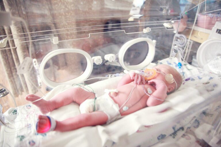
For the baby, the main issue is planned earlier birth, increasing the chance of prematurity and NICU care.
Emotionally, some parents experience anxiety, depressive symptoms, or post-traumatic stress symptoms after a high-risk birth or major surgery. If intrusive memories, panic, or persistent low mood appear, bring it up early—support is part of recovery.
Care planning once placenta accreta is suspected
Why a multidisciplinary team is used
PAS planning often involves maternal–fetal medicine, experienced obstetric surgeons, anesthesia, neonatology, and a blood bank, interventional radiology and urology may join depending on severity.
Blood planning and anemia treatment
Teams may prepare a massive transfusion protocol, crossmatched blood, and sometimes cell salvage. Iron deficiency/anemia is treated when there is time (oral or IV iron) to improve reserves.
Practical coordination
Expect more appointments and potentially travel. Many families benefit from choosing one team contact person and planning childcare/work leave early.
Planning delivery with placenta accreta
Many protocols plan birth around 34–35+6 weeks in higher-risk cases (some centers extend to 37 weeks for selected stable situations). Antenatal corticosteroids may be offered for fetal lung maturation if preterm delivery is planned.
A planned cesarean is often preferred, particularly with placenta previa, to avoid labor-related bleeding. Surgeons map placental location to reduce the chance of cutting through the placenta.
Preparation commonly includes two large-bore IV lines, close blood pressure monitoring (sometimes an arterial line), and immediate access to blood products.
Interventional radiology options (balloon occlusion/embolization) are used in some hospitals, evidence is mixed and practice varies.
Standard surgical approach: cesarean hysterectomy
A cesarean hysterectomy means delivering the baby by cesarean and then removing the uterus during the same operation. In many cases of placenta accreta, it is the safest definitive way to control bleeding.
Often the placenta is left in situ (not peeled away). Removing the uterus with the placenta still attached can reduce catastrophic hemorrhage.
Conservative (fertility-sparing) options: carefully selected
In specific situations, uterus-sparing strategies may be discussed, such as leaving the placenta in situ with close follow-up, or focal resection with uterine repair when invasion is limited. These approaches can carry higher risks of delayed hemorrhage, infection/sepsis, repeat procedures, and delayed hysterectomy.
Methotrexate is not routinely used by many specialist teams due to limited biologic rationale late in pregnancy and potential toxicity.
Postpartum recovery and follow-up
After surgery for placenta accreta, monitoring may occur in ICU/high-dependency units for 24–48 hours, with close checks of bleeding, urine output, vital signs, and labs (hemoglobin, platelets, clotting tests, fibrinogen).
Pain relief is typically multimodal, and early assisted mobility lowers complication risk. Because clot risk is higher after major pelvic surgery, compression devices and sometimes low-molecular-weight heparin are used once bleeding is controlled.
If bladder repair was needed, catheter care may be longer, with attention to urinary symptoms.
Feeding can be challenging if the baby is in NICU, early lactation support and pumping guidance can help.
If the placenta was left inside the uterus, follow-up imaging and clear warning signs are essential (fever, heavy bleeding, worsening pain, new urinary symptoms).
Life after placenta accreta and future pregnancies
After hysterectomy, recovery follows major pelvic surgery pathways and fertility ends. After uterus-sparing care, recovery can be longer and less predictable.
If the uterus is preserved, a future pregnancy is possible but high risk. Recurrence after conservative management has been reported around 15–30% across studies, so early targeted ultrasound is usually planned.
Key takeaways
- Placenta accreta is abnormal deep attachment of the placenta and can cause severe bleeding at delivery.
- The strongest risk factor for placenta accreta is a prior cesarean, especially with placenta previa.
- Many parents have no symptoms, any third-trimester bleeding needs prompt obstetric advice.
- Ultrasound with Doppler is first-line, MRI helps in selected cases.
- Planned birth in an experienced center with transfusion readiness and a coordinated team improves safety.
- Common definitive treatment is planned cesarean hysterectomy with the placenta left in situ.
- Conservative options exist for selected cases but require close monitoring and accept higher risks.
- Professionals can support medical, feeding, and emotional recovery, for tailored guidance and free child health questionnaires, parents can download the Heloa app.
Questions Parents Ask
Can placenta accreta be missed on ultrasound?
Yes, it can happen—especially if the placenta is posterior (on the back wall), if imaging windows are limited, or if signs are subtle early on. Rassurez-vous: targeted ultrasounds by an experienced team, sometimes repeated over time, improve detection. In selected situations, an MRI may be offered to clarify how deep the placenta attaches and whether nearby organs could be involved.
What is the survival rate with placenta accreta?
Most parents do well when placenta accreta is anticipated and delivery is planned in a specialist center with blood products immediately available. The condition is serious mainly because of bleeding risk, not because it is “untreatable.” Outcomes are generally best with coordinated care (experienced obstetric surgeons, anesthesia, blood bank, NICU), and many families leave hospital safe—though recovery can take time, both physically and emotionally.
If I want more children, is hysterectomy always the only option?
Not always. A planned cesarean hysterectomy is often the safest definitive approach, but in carefully selected cases, conservative (uterus-sparing) management may be discussed. It’s important to know these options can come with higher chances of delayed bleeding, infection, and later emergency surgery. If fertility is a priority, you can ask about your individual risk level, follow-up plan, and what a future pregnancy would involve (often early specialist scans and close monitoring).

Further reading :