You brought a baby into the world, now what about the placenta. Most of the time it follows within minutes, almost like the last step in a well rehearsed sequence. Sometimes it does not. When the placenta stays inside the uterus beyond the usual window, clinicians call it retained placenta. You might wonder, is it dangerous, how would I notice it, what would my team do, and how could this affect recovery and future pregnancies. The short version, timing guides action, heavy bleeding triggers faster steps, and with planned care most parents do well. The long version, explained clearly and kindly, starts here.
What retained placenta means and why timing matters
Retained placenta means some or all of the placenta remains attached or trapped after the baby is born. Many teams watch a practical clock, about 30 minutes is a common threshold, and in well monitored situations with stable vitals and light bleeding, some teams may wait up to 45 to 60 minutes. If bleeding is heavy, they escalate sooner. While tissue remains inside, the uterus cannot squeeze blood vessels closed effectively, bleeding rises, and infection risk increases. That is why retained placenta gets prompt attention.
Two patterns show up in real life:
- Complete non delivery, the entire placenta has not come out
- Retained fragments, most is delivered but small pieces or membranes remain
You might ask, would I feel this. Often the first sign is bleeding that keeps going or gets heavier, sometimes with clots, sometimes with dizziness or a racing pulse.


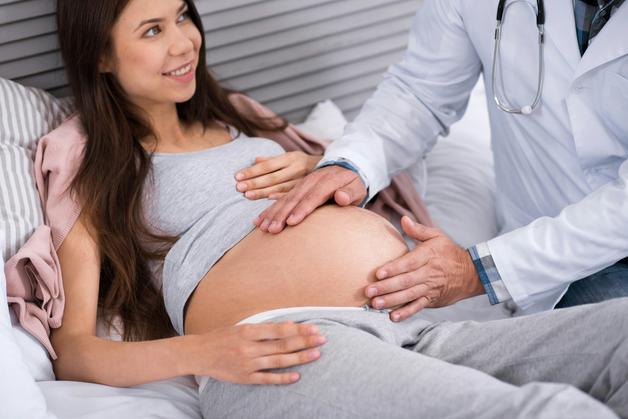
The third stage of labor, a quick tour
The uterus is a powerful muscle. After the baby is born, strong contractions deform the inner lining, reduce blood flow between uterus and placenta, and shear the placenta off the uterine wall. With contractions and controlled cord traction, the placenta slides down and out. Then the same contractions compress open vessels like a firm hand wringing a soaked cloth, limiting blood loss. In straightforward births this takes 10 to 20 minutes.
Parents sometimes ask, can a hands off approach be safe. Many places use active management of the third stage of labor, which means giving a medicine to contract the uterus, guiding the cord gently, and checking the placenta to ensure it is complete. This approach lowers the chances of heavy bleeding and retained tissue.
Types of retained placenta
Trapped placenta
The placenta separates but cannot pass through because the cervix closes or a constriction ring forms. Bleeding can continue, the uterus may cramp hard, and removal may require a procedure under anesthesia. Quick resolution limits bleeding and infection.
Adherent placenta, placenta adherens
Separation is incomplete because the normal cleavage plane is thin or scarred. The placenta stays stuck and manual removal is often needed. This is distinct from Placenta accreta spectrum, where the attachment goes deeper into or through the muscle.
Placenta accreta spectrum
Attachment is abnormally deep, ranging from accreta, firmly stuck on the muscle surface, to increta, into the muscle, to percreta, through the wall and possibly into nearby organs. Prior cesarean birth, uterine surgery, and placenta previa raise risk. When suspected, teams plan delivery in a center with surgical support, blood bank, and interventional radiology, sometimes with a hysterectomy if bleeding cannot be controlled.
Retained placental fragments and membranes
Small pieces can remain after most of the placenta delivers. Symptoms may appear days to weeks later, new heavy bleeding after improvement, pelvic pain, or a foul smell. Ultrasound confirms the diagnosis, then targeted removal with suction curettage or hysteroscopy is common, with antibiotics if infection is suspected.
How common retained placenta is
Frequency varies by definition and practice patterns, but retained placenta is uncommon. Using the 30 minute window, studies often report 0.1 to 3 percent after vaginal birth. Active third stage care lowers the chance.
Why retained placenta happens, mechanisms in plain language
Most cases trace back to three mechanisms:
- Failure of clean separation, the decidual layer is thin or scarred, sometimes related to prior surgery
- Trapping above a closing cervix, the placenta is free but cannot exit
- Poor uterine tone, called uterine atony, the muscle is too soft to detach or expel tissue and cannot clamp vessels
Clinicians integrate history, how the uterus feels, firm versus boggy, bleeding pattern, and imaging to decide next steps.
Risk factors that raise attention
History and anatomy
- Prior retained placenta
- Previous cesarean, myomectomy, or repeated curettage
- Large fibroids, uterine anomalies, advanced maternal age, higher parity
- Conception with IVF or ART in some studies
Pregnancy and birth context
- Placenta previa or low lying placenta
- Preterm birth or stillbirth
- Very long or very fast labor, induction or augmentation, chorioamnionitis, operative vaginal birth
- Early or forceful traction on the cord with tearing of placental tissue
System and preparation
- Treating anemia increases resilience to blood loss
- Ready access to uterotonics, blood products, trained staff, and clear escalation pathways improves outcomes
Risk factors inform planning, they do not predict the future with certainty.
Signs and symptoms parents can recognize
Immediate warning signs after birth
- Placenta not delivered within the expected time frame
- Heavy bleeding or large clots, soaking pads quickly
- A soft boggy uterus that does not stay firm with massage and medicine, or a very tense and painful uterus
- Dizziness, pale skin, low blood pressure, fast pulse
Delayed warning signs days to weeks later
- New heavy bleeding after earlier improvement, called secondary postpartum hemorrhage (PPH)
- Fever, pelvic pain, foul smelling discharge
- The uterus seems larger than expected with prolonged lochia
After cesarean birth, difficulty removing the placenta during surgery, persistent bleeding, fever, or prolonged lochia can point to retained tissue or abnormal placentation.
Seek urgent care for heavy bleeding, faintness, severe pain, or fever.
How clinicians diagnose retained placenta
Bedside assessment
- Time since birth, bleeding amount, and uterine tone guide decisions
- The delivered placenta and membranes are inspected for missing lobes or torn edges
Imaging
- Ultrasound of the uterus looks for echogenic tissue in the cavity
- transvaginal ultrasound gives detailed views, Doppler may show internal blood flow within retained tissue
Laboratory tests
- Hemoglobin and hematocrit to estimate blood loss
- Platelets and coagulation tests when bleeding is significant
- Cultures and inflammatory markers when infection is suspected
Differential diagnosis
- Uterine atony without retained tissue
- Retained membranes only, often prolonged lighter bleeding
- Intrauterine hematoma, pooled blood inside the uterus
- Bleeding related to clotting disorders
First steps in the delivery room
Priorities are simple and life saving. Stabilize the parent, place IV access, start fluids, give oxygen if needed, and monitor vitals. Estimate blood loss, prepare the team, and use uterotonics promptly. oxytocin is first line to strengthen contractions. If bleeding is heavy, clinicians may use tranexamic acid early to support clot stability.
Practical measures
- External uterine massage
- Make sure the bladder is empty
- Try controlled traction once the uterus is firm
- If the placenta will not deliver, or if bleeding continues, move to the operating room for manual removal under anesthesia
Treatment options and what to expect
Manual removal of the placenta
- Indicated when the expected time has passed or when bleeding is ongoing
- Performed under regional or general anesthesia
- The clinician separates the placenta from the uterine wall, removes clots and fragments, and checks that the cavity is clear
Curettage and targeted removal
- If imaging or exam suggests residual fragments, suction curettage may follow
- Persistent fragments can be removed with hysteroscopy, a thin camera inside the uterus that allows precise removal, with a lower adhesion risk in experienced hands
Bleeding control and supportive care
- Intrauterine balloon tamponade, for example a Bakri balloon, applies pressure from within the uterus
- Compression sutures in the operating room can limit bleeding while preserving the uterus
- Interventional radiology can perform uterine artery embolization to reduce blood flow
- Transfusion and blood products are given as needed
Definitive surgery
- In rare and severe cases, especially with Placenta accreta spectrum, hysterectomy may be the safest option if bleeding is uncontrollable
Antibiotics and infection control
- Prophylactic antibiotics are common around invasive procedures
- If signs of endometritis appear, fever, pelvic pain, foul discharge, targeted antibiotics begin promptly
Fertility notes
- Most parents retain fertility after a retained placenta, especially with timely care
- Repeated curettage or severe infection can create intrauterine adhesions, which may need treatment before a future pregnancy
Special situations
Trapped placenta
- The placenta has separated but is incarcerated above a closing cervix
- Specific maneuvers under anesthesia and short acting relaxation medicines may be used, followed by uterotonics
Adherent placenta versus accreta spectrum
- Adherent placenta usually separates with manual techniques
- Accreta spectrum calls for a different plan, often in a tertiary center with a multidisciplinary team
Preterm birth, multiples, stillbirth
- Smaller or fragile placentas behave differently, and the team balances gentle techniques with safety
- Emotional care matters, ask for a debrief and support
Cesarean birth
- The surgeon removes the placenta directly, so classic retained placenta is less common
- Abnormal invasion patterns can complicate removal and require advanced planning
Rh negative parents
- If the baby is Rh positive, anti D immunoglobulin is given after birth or after invasive procedures to protect future pregnancies
Recovery, aftercare, and planning the next pregnancy
Monitoring and home care
- In hospital, teams check bleeding, vitals, uterine tone, and pain
- At home, call for heavy bleeding, large clots, fever or chills, foul discharge, severe pelvic pain, marked dizziness, shortness of breath, or extreme fatigue
Tests and medicines
- Follow up ultrasound can confirm the uterus is clear
- Blood tests help track anemia, iron replacement by mouth or IV is common after significant loss
- Pain relief with acetaminophen or ibuprofen is typically compatible with breastfeeding
Breastfeeding and mental well being
- Breastfeeding can continue, even after procedures and antibiotics in most cases
- Processing the experience helps, ask your team for a debrief and support options
Future pregnancy planning
- A follow up visit reviews what happened, likely causes, and any scarring
- Next time, care may include early scans to check placental position and attachment, with a birth plan tailored to your risk profile
When to seek medical help after birth
Call promptly if you experience:
- Heavy bleeding that soaks pads quickly or large clots
- Fainting, severe dizziness, or feeling very unwell
- High fever, severe pelvic or abdominal pain, or foul smelling discharge
Normal lochia lightens over two to six weeks. New heavy bleeding after earlier improvement, persistent heavy flow, fever, foul discharge, or severe cramping deserves evaluation.
Key takeaways
- Retained placenta means placental tissue remains in the uterus after birth, the typical watch window is about 30 minutes, longer if stable, shorter if bleeding is heavy.
- Main drivers include incomplete separation, trapping above a closing cervix, and uterine atony that prevents the uterus from clamping down.
- Warning signs include heavy bleeding, a boggy uterus, dizziness, fever, pelvic pain, and foul discharge.
- Diagnosis relies on clinical assessment, ultrasound including transvaginal ultrasound, sometimes with Doppler, and blood tests.
- Treatments include uterotonics like oxytocin, manual removal, suction curettage, targeted hysteroscopy, balloon tamponade with a Bakri balloon, and escalation to surgery for severe cases or Placenta accreta spectrum.
- Infection such as endometritis is managed with antibiotics, and bleeding is supported with measures like tranexamic acid and transfusion when needed.
- Most parents recover well and retain fertility, with planning for a future pregnancy based on what happened this time.
- Support exists at every step, your care team can personalize decisions to your situation, and you can use the Heloa app for trustworthy guidance. Download the application Heloa for personalized tips and free child health questionnaires.
Retained placenta deserves prompt recognition and calm, organized care. With the right plan and timely support, recovery is the rule, and confidence for future births grows.
Questions Parents Ask
Can a retained placenta resolve on its own?
Yes — sometimes. When bleeding is light and the parent is stable, teams commonly watch for about 30 minutes and in some settings up to 45–60 minutes because the placenta can separate and pass on its own. However, the longer tissue stays inside, the greater the chance of heavier bleeding or infection, so clinicians will intervene sooner if bleeding increases or vital signs change. Small fragments may also be missed and only cause symptoms days to weeks later; in that case ultrasound and targeted removal are often needed. It’s natural to worry — ask your care team how long they plan to observe and what signs would prompt treatment.
What is manual removal of the placenta like?
Manual removal is usually done in the operating room with pain relief and monitoring. With regional (spinal/epidural) or general anesthesia, the clinician gently inserts a gloved hand into the uterus, separates the placenta from the lining, and removes any clots or fragments. The procedure is typically brief; afterwards the team makes sure bleeding is controlled and may perform suction curettage if needed. Expect close observation for a few hours, pain medication as needed, and information about bleeding and infection signs before discharge. Many parents can breastfeed soon afterwards, and staff typically offer emotional support and a debrief once you’re recovering.
What anesthesia options are available and what should I expect?
Options depend on your situation and how urgent treatment is. If you already have a working epidural or spinal, it can usually be topped up so you remain awake but numb — a common choice for manual removal. If there’s heavy bleeding, rapid intervention, or other concerns, general anesthesia (put to sleep) may be used so the team can work quickly and safely. Regional anesthesia avoids the grogginess of general anesthesia and lets you be present for immediate postpartum moments, while general anesthesia requires a short recovery period and sometimes temporary separation from the baby. Your care team can explain risks and benefits and try to align the plan with your preferences whenever the clinical situation allows.

Further reading: