You open the report, you see a number, your mind races. Is a 12th percentile okay, what about a 90th, should the next scan happen sooner. Take a breath. A fetal growth percentile is a position on a curve, not a verdict. It helps the team and you talk clearly about size, trend, and wellbeing. You will see how the number is calculated, how it is tracked across weeks, and how it fits with other checks like blood flow and amniotic fluid. Most importantly, you will learn what matters right now, the trajectory, the context, and the plan that follows.
Fetal growth percentile, how to read it and why it helps
A percentile describes where a measurement sits compared with babies at the same week of pregnancy. The 50th percentile means middle of the road, half smaller, half larger. The 10th means smaller than most for that week, the 90th means larger than most.
- A percentile is often reported for an Estimated Fetal Weight, also called EFW, or for a single measure such as the abdominal circumference. You may also see a z-score, a number of standard deviations from the median, useful to quantify how far from average the point lies.
- One point is a snapshot. The path across time, the growth line from scan to scan, that is what changes decisions.
- Most babies between the 10th and 90th fetal growth percentile do well, especially if growth stays steady and the rest of the assessment is reassuring.
You may wonder, is a single low fetal growth percentile always a problem. Not always. Small parents often grow smaller babies who are healthy. Is a very high fetal growth percentile always a problem. Not by itself, but it triggers checks for glucose metabolism and birth planning.
What clinicians look at beyond the number
A fetal growth percentile is a starting point. The team layers clinical context and tests that check placental function and fetal reserve.


- Screening for restriction and for large size, often labeled as SGA small for gestational age or LGA large for gestational age
- The trend in size, the growth velocity, and whether centiles are crossed downward or upward
- Blood flow assessment with Doppler ultrasound, fetal heart rate patterns, and fluid volume
- Timing of follow up scans and whether to add extra tests or change birth planning
How size is measured and converted to a fetal growth percentile
Ultrasound biometry and inputs
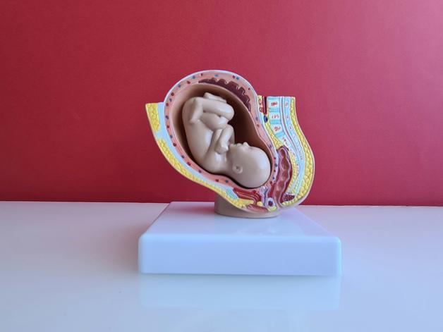
The sonographer measures standard anatomical planes to estimate size. Accuracy comes from technique and clear images.
- Core measurements, BPD biparietal diameter, HC head circumference, AC abdominal circumference, FL femur length
- Why each matters, the abdomen reflects reserves and is highly influential on weight estimates, head measures follow brain growth, femur length reflects skeletal growth
- These inputs are combined to calculate EFW, then the EFW is located on a curve to create a fetal growth percentile
You might see the measurement set on your report. Ask for the raw numbers in millimeters and the formula used. This helps create a consistent record across visits with the same reference.
Gestational dating, the foundation of every percentile
All percentiles are tied to gestational age. If the dating is off, a normal baby can appear small or large.
- Early dating with crown rump length is usually the most accurate approach
- If a scan shows an unexpected fetal growth percentile, clinicians often confirm dating first before labeling a growth problem
From measurements to EFW
Established formulas convert biometry to a weight estimate.
- The Hadlock formula family is widely used, combining different sets of BPD, HC, AC, FL
- The Shepard method is older, you may still see it in some reports
- In late pregnancy, when parts are harder to see, teams sometimes rely more on AC centered estimates
About uncertainty, no estimate is perfect. Typical EFW error is about 10 to 15 percent, and can reach 20 percent late in pregnancy. Image quality, fetal position, and operator skill all matter. Results near a threshold, such as the ninth to eleventh fetal growth percentile, are often rechecked with a short interval follow up.
From EFW to percentiles and growth charts
Your EFW, or sometimes a single measure like AC, is compared with a reference distribution for the exact week.
- References you may see, INTERGROWTH 21st growth standards, WHO fetal growth standards, Hadlock reference curves, population specific charts, and customized growth charts that adjust for maternal size and fetal sex
- Some clinics use sex specific charts, others use a single curve for both
Parent tip, note the reference used on your report and try to keep it consistent across visits. A switch to a different reference can shift the reported fetal growth percentile even if your baby grew as expected.
Ultrasound best practices and common errors
High quality biometry depends on standard technique and repetition.
- The sonographer uses correct planes, places calipers on outer bony edges for skull and on skin line for abdomen, and measures the femur on its longest axis
- Measurements are often repeated then averaged, this smooths random error
- Keep scans at the same center when possible, this reduces variation between machines and operators
- Ask that the report lists raw measurements, EFW formula, and the growth chart applied
If a percentile drops and the baby was curled tightly or the images were grainy, a repeat with the same experienced operator can change the point. It is reasonable to ask whether multiple measures were averaged.
Interpreting patterns across pregnancy
Trimester specific expectations
- First trimester, best for dating with crown rump length, size is less helpful to label growth problems
- Second trimester, measurements become more reliable for tracking growth, many baseline percentiles are established here
- Third trimester, focus shifts to trend and wellbeing, Dopplers and growth velocity guide decisions
Thresholds and what they usually mean
- Below the third, higher concern for restriction, closer monitoring and tests, particularly if Dopplers are abnormal
- Third to tenth, can be constitutional smallness or mild restriction, surveillance usually increases
- Tenth to ninetieth, common range, most babies here do well
- Ninetieth to ninety seventh, larger than average, check for gestational diabetes and discuss delivery planning if the trend persists
- Above the ninety seventh, higher chance of macrosomia, attention to maternal glucose, monitoring, and birth planning
Static percentile versus growth velocity
A static point is one moment. Movement across the chart reveals biology. A steady line at a low fetal growth percentile can be acceptable, a sharp drop across centiles is more concerning and often prompts added Dopplers and shorter intervals between scans.
Discordant parameters and constitutional variation
Isolated small AC with normal HC and FL can be normal variation or early restriction. Head sparing, the head stays on track while the abdomen lags, may reflect placental stress and a brain sparing effect. Teams tend to add umbilical artery Doppler, middle cerebral artery Doppler, and short interval follow up to clarify the trend.
Reading a growth chart without getting lost
- The horizontal axis shows gestational weeks, the vertical axis shows a measurement like EFW in grams or a circumference in millimeters
- Lines show centiles, for example 2.5, 5, 10, 25, 50, 75, 90, 95, 97.5
- Your baby’s estimate is plotted on the curve to read the fetal growth percentile
Indicative EFW values, Hadlock based, approximate, typical error 10 to 15 percent
- 24 weeks, 10th about 576 g, 50th about 665 g, 90th about 765 g
- 28 weeks, 10th about 1026 g, 50th about 1189 g, 90th about 1368 g
- 32 weeks, 10th about 1635 g, 50th about 1901 g, 90th about 2187 g
- 36 weeks, 10th about 2352 g, 50th about 2745 g, 90th about 3153 g
- 40 weeks, 10th about 3084 g, 50th about 3617 g, 90th about 4135 g
Three mini cases to orient your expectations
Case 1, 28 weeks, EFW 1050 g, around the 12th percentile
Interpretation, close to the 10th, plan a follow up in 2 to 4 weeks to check the trajectory. Next steps, consider risk factors like smoking or high blood pressure, add Dopplers if the curve dips.Case 2, 32 weeks, EFW 2100 g, around the 85th percentile
Interpretation, high zone but not alarming. Next steps, ensure diabetes screening is current, monitor growth, begin conversations about birth planning if size continues to trend high.Case 3, 38 weeks, EFW 4000 g, above the 90th percentile
Interpretation, suspected large size. Next steps, maternal glucose assessment if not done, discuss induction timing or delivery mode based on the broader clinical picture.
Practical classifications, why labels appear on reports
SGA small for gestational age, EFW below the 10th percentile
This is a statistical category used to guide surveillance. Some babies are constitutionally small and healthy.LGA large for gestational age, EFW above the 90th percentile
This points to a larger size and a higher chance of shoulder difficulty at birth, planning reduces risk.
These labels direct appropriate monitoring. They do not determine your baby’s future.
SGA and LGA, causes, risks, and follow up
SGA, possible causes include placental insufficiency, high blood pressure, smoking, inadequate nutrition, infections, fetal anomalies, and genetic variation. Risks include temperature instability and low blood sugar after birth. Follow up often includes serial growth scans every 2 to 4 weeks, uterine artery Doppler, umbilical artery Doppler, and maternal assessment. Hospital care can be needed if blood flow is severely abnormal or distress is suspected.
LGA, common drivers include gestational diabetes, higher BMI, prior large infant, and family factors. Risks include shoulder dystocia, a higher cesarean rate, and neonatal low blood sugar if maternal diabetes is present. Follow up includes an oral glucose tolerance test when indicated, glucose management, and a closer eye on growth and delivery planning.
Factors that can shift the fetal growth percentile
- Maternal characteristics, height, weight, prior history, age, diabetes, high blood pressure
- Lifestyle, smoking, alcohol, nutrition, activity level
- Conditions, preeclampsia, infections, chromosomal anomalies
- Technical, dating accuracy, image quality, operator experience, chosen reference chart
Additional tests and when plans change
When a fetal growth percentile sits near the borders, or when the trend dips or surges, follow up usually intensifies in a tailored way.
- Serial growth scan every 2 to 4 weeks near the 10th or 90th
- Doppler ultrasound of uteroplacental circulation, absent or reversed end diastolic flow is a severity sign that can lead to hospital monitoring or early birth
- Fetal tests as indicated, nonstress testing, abbreviated as NST, biophysical profile, and fluid checks such as amniotic fluid index
- Maternal tests, oral glucose tolerance for diabetes, iron and nutrition checks as needed
Fluid volume adds context. Low fluid, oligohydramnios, and high fluid, polyhydramnios, provide additional clues and can shift timing decisions.
When to discuss induction or a cesarean
Decisions blend the fetal growth percentile with the entire clinical picture. Examples that prompt a planned birth include markedly abnormal Dopplers, suspected fetal distress, or very high EFW with poorly controlled diabetes. Thresholds are individualized, benefits and risks are explained, and plans are written down after shared decision making.
Impact on labor and newborn care
- Low percentile, closer surveillance, regular Dopplers, and preparation for an earlier birth if needed, with the neonatal team ready
- High percentile, attention to maternal glucose, possible induction around 38 to 39 weeks in the setting of diabetes, and a clear plan for mode of birth
During labor, small babies have a higher risk of intolerance to contractions, the team adapts in real time. Large babies have a higher risk of shoulder dystocia and postpartum bleeding, preparation and prompt action reduce these risks.
After birth, small babies get early checks for blood sugar and temperature, larger babies born to diabetic parents receive early glucose monitoring to prevent and manage hypoglycemia.
Practical ways to support healthy growth
- Nutrition, for suspected SGA, optimize intake, correct deficiencies, stop smoking, and limit alcohol. For LGA with diabetes, seek dietitian guidance, balance glucose with food and medication if prescribed.
- Activity, stay active with pregnancy safe exercise if your clinician approves
- Continuity, record scan dates, raw measures, EFW in grams, the reference used, and keep your reports so trend interpretation stays consistent
Red flags that merit prompt contact with your maternity unit, a marked drop in fetal movements, fluid loss, bleeding, severe pain, fever.
Useful tools to make sense of the numbers
Ask for a complete report, raw measures, EFW in grams, fetal growth percentile, reference chart, date, and operator. Validated calculators that incorporate Hadlock or INTERGROWTH 21st growth standards help place the percentile and track the trajectory. Pair biometric data with WHO fetal growth standards or local references consistently, this reduces confusion and supports clear plans.
References, nuance, and balance
Growth curves are statistical models, your baby is an individual. The fetal growth percentile is a shared language that guides proportionate care. Uncertainty is part of every estimate, which is why trend and context matter more than a single point. When something feels unclear, ask the team to walk you through the graph, the blood flow findings, and the next milestone.
Key takeaways
- A fetal growth percentile places your baby on a curve for a given week. The 10th to 90th range is common and usually reassuring when growth is steady.
- EFW comes from fetal biometry, then is converted to an EFW percentile using references like the Hadlock family, INTERGROWTH 21st growth standards, or WHO fetal growth standards.
- Trend beats a snapshot. Near threshold results are usually confirmed with a short interval follow up, especially when measurement uncertainty is high.
- Labels such as SGA small for gestational age and LGA large for gestational age organize care, they do not predict a lifetime trajectory.
- Blood flow testing with Doppler ultrasound, including umbilical artery Doppler and uterine artery Doppler, plus fluid checks like amniotic fluid index, round out the picture.
- Birth planning considers percentile, Dopplers, fluid, maternal health, and growth velocity, the plan is individualized and revisited as new data arrive.
- Support is available. For personalized guidance and free child health questionnaires, you can download the Heloa app, https://app.adjust.com/1g586ft8
Throughout your pregnancy, lean on clear data, steady follow up, and open conversations. If a fetal growth percentile raises questions, ask them, your care team will use science and empathy to map the next step.
Questions Parents Ask
How do I use an online fetal growth percentile calculator?
Most calculators ask for either an estimated fetal weight (EFW) in grams or the raw ultrasound measures (BPD, HC, AC, FL) plus the gestational age. Enter the exact numbers from your report, pick the growth reference the tool uses (Hadlock, INTERGROWTH, WHO, etc.), and submit to get a percentile. A few friendly reminders: calculators are only as good as the inputs and the chart they use, so try to use the same reference over time; note the reported margin of error; and keep a copy of the raw measures and which chart was used. If anything feels unclear, bring the printout to your clinician for a quick walk-through — they can interpret the result in context.
What is the NICHD fetal growth calculator and how does it differ from other charts?
The NICHD (National Institute of Child Health and Human Development) fetal growth work produced reference curves based on U.S. study data and highlighted differences across populations. Unlike INTERGROWTH-21st (an international prescriptive standard) or older Hadlock curves, NICHD data account for population variation seen in the study cohorts. Different charts can shift percentiles slightly; none are perfect. What matters most is consistent use of a chosen chart and interpreting percentiles together with trend, Dopplers, fluid, and maternal health.
Can I trust online calculators — are their percentiles accurate?
Online tools are useful for orientation but are not a medical verdict. They reliably translate your numbers into a percentile for the selected chart, but several limits remain: EFW itself has typical ultrasound error (often 10–15% or more late in pregnancy), different charts give different percentiles, and operator or dating inaccuracies can change results. Treat calculator output as a conversation starter rather than a final answer. If a result is near a threshold or worries you, ask for the raw measurements, the chart used, and a clinician’s interpretation — that combination gives the clearest next step.

Further reading :