You have a decision to make, and it is not a small one. Amniocentesis can bring clarity when screening has raised a question, when ultrasound shows something unexpected, or when a family condition needs a yes or no answer. You may be wondering, how is the sample taken, what does it actually test, how safe is it, and how long before we know more. The short version, a carefully guided needle collects a small amount of fluid from around the baby, the lab studies fetal cells and molecules, and the results can confirm many genetic or chromosomal findings that screening only estimates.
What amniocentesis is and why it is offered
Amniocentesis is a diagnostic test, not a screening test. A trained clinician collects amniotic fluid, which contains fetal cells and signaling molecules, then a laboratory evaluates chromosomes, specific genes, and sometimes infectious agents. The goal is prenatal diagnosis, which means moving from a risk estimate to a confirmed answer.
In one breath definition, amniocentesis is an ultrasound guided procedure that removes a small volume of amniotic fluid so that laboratories can run a fetal karyotype, chromosomal analysis, chromosomal microarray, and other targeted assays to detect chromosomal differences, selected genetic conditions, and some infections.
Why consider it


- A high risk screen on noninvasive prenatal testing or serum screening
- An ultrasound finding that needs genetic confirmation
- A known familial condition such as cystic fibrosis or spinal muscular atrophy
- A parental chromosomal rearrangement, for example a balanced translocation
- Concern for infection such as cytomegalovirus, when results would change care
You might ask, will it find everything. No test can. Amniocentesis answers a defined question, and the answer depends on the specific analysis ordered.
How the procedure works, what you will feel
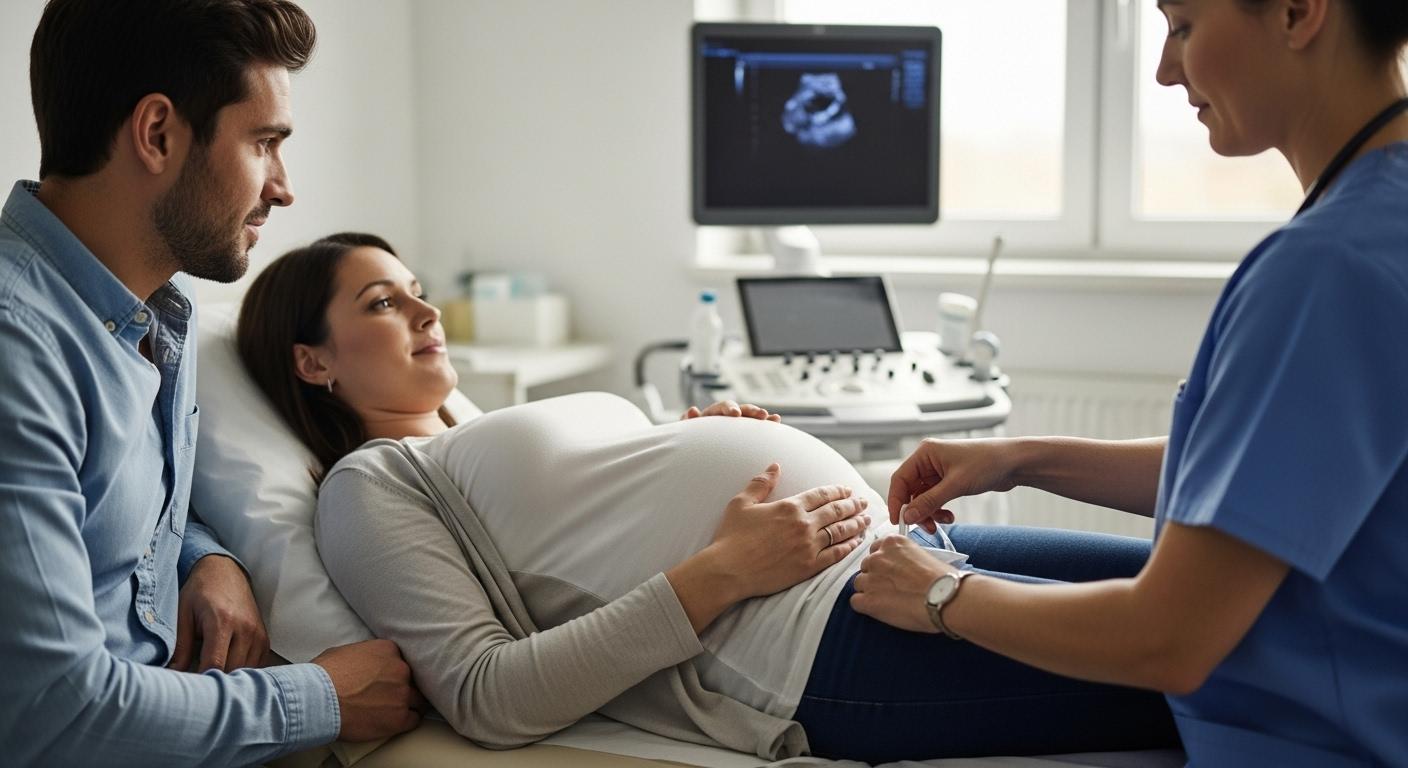
The team uses ultrasound guided amniocentesis to map the placenta and baby, then chooses a safe pocket of fluid. After sterile skin preparation, a fine needle passes through the abdomen and into the amniotic sac. The sampling of about 15 to 30 milliliters usually takes under one minute. The entire visit often fits into 10 to 30 minutes, depending on fetal position.
What does it feel like
- Most people describe a brief pinch or pressure
- Mild cramping can follow and usually settles within a day
- Local anesthetic can be used depending on the center
A practical note, calm breathing, a comfortable position, and a clear explanation from the team make a real difference.
When amniocentesis is recommended
Common indications
- Positive or high risk result on cell free DNA testing, first trimester or second trimester serum screening
- Ultrasound markers that raise concern and require confirmation
- Advanced maternal age with individualized discussion
- Family history of a single gene condition or a known chromosomal rearrangement
Examples of conditions assessed
- Trisomy 21, trisomy 18, trisomy 13
- Sex chromosome differences such as Turner syndrome
- Submicroscopic deletions or duplications, also called copy number variants
- Targeted single gene testing if a familial variant is known
- When indicated, infectious testing for CMV or toxoplasma
Values matter. Think about what you want to know now, how answers might change your pregnancy care or delivery planning, and what level of uncertainty you are comfortable carrying. This is where genetic counseling shines, both before testing and after results.
Timing and types of amniocentesis
Second trimester timing
The optimal window is 15 to 20 weeks, often around 16 to 18 weeks. There is enough fluid to sample, cell yield is strong, and safety is favorable.
Early and late procedures
Early amniocentesis, around 11 to 14 weeks, is less common because complication rates are higher than with chorionic villus sampling at that same stage. In the third trimester, amniocentesis is uncommon but may be used to evaluate fetal infection, to address a pressing diagnostic question, or to assess markers of lung maturity when early delivery is considered.
Special scenarios
- Twins, separate samples may be taken for each sac with careful labeling
- Anterior placenta or uterine fibroids, the needle path is adjusted using real time ultrasound
- Therapeutic procedures like amnioreduction are different from diagnostic amniocentesis, the goals and risks are not the same
Step by step, before, during, and after
Before the procedure
- Review obstetric and family history
- Confirm blood group and Rh status, give anti D immune globulin after the procedure if indicated
- Share prior results from noninvasive prenatal testing, serum screening, and ultrasound
- Discuss medicines such as anticoagulants or high dose aspirin
- Sign informed consent covering goals, limits, alternatives, and risks
Day of practical checklist
- Bring your blood group card and medication list
- Wear clothing that allows easy abdominal access
- Hydrate and plan for quiet time afterward
- Write down your key questions
During the procedure
Ultrasound maps fetal and placental positions. After sterile preparation, the needle enters the amniotic sac away from the fetus and placenta, the clinician aspirates the planned volume, and the sample goes to the lab in labeled tubes matched to the required assays.
After the procedure
- Expect mild cramping or light spotting for 24 to 48 hours
- Most people rest for a day and then resume light activity
- Your team will share warning signs and a direct contact number
What amniocentesis tests for
Chromosomes and the genome
- Karyotype, a global look at chromosome number and structure to detect trisomies and large rearrangements
- chromosomal microarray, higher resolution analysis to detect copy number variants that a karyotype can miss
- rapid aneuploidy testing, such as QF PCR or FISH, early answers within two to three days for the most common aneuploidies
Targeted molecular analysis
- If a familial variant is known, the lab can check for that exact change by PCR or sequencing
- Broader sequencing can be considered in specific contexts, with careful pre test counseling about potential findings
Neural tube defects and infection
- Amniotic fluid AFP and acetylcholinesterase help assess open neural tube defects alongside ultrasound
- For suspected infection, PCR can test for CMV or toxoplasma, particularly when results would change care
Accuracy, limits, and turnaround times
How accurate are the tests
Rapid assays are highly accurate for common trisomies. Karyotype evaluates the full set of chromosomes. Microarray increases the chance of finding submicroscopic deletions or duplications. Targeted testing for a known familial variant is very precise.
Limits to keep in mind
- Mosaicism can complicate interpretation because different tissues may show different cell lines
- Maternal cell contamination can affect results, laboratories use methods to detect and reduce it
- Not every condition is detectable, especially if a gene is not specifically tested or if the disorder is metabolic and not visible on chromosomal studies
- Positive rapid results are usually confirmed by full karyotype or microarray, parental studies may clarify whether a detected variant is inherited
Turnaround times
- Rapid QF PCR or FISH, about 48 to 72 hours
- Karyotype with culture, about 2 to 3 weeks
- Microarray, often 3 to 4 weeks
- Infectious PCR, from a few days to two weeks depending on the laboratory
Risks, complications, and warning signs
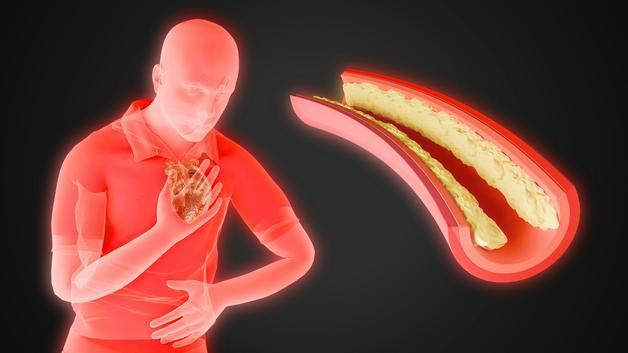
Miscarriage risk
Large series estimate a procedure related risk from about 0.3 percent to 1 percent. Most of this risk is concentrated in the first days up to about two weeks after sampling. Operator experience, continuous ultrasound guidance, and the clinical scenario influence that risk.
Other potential effects
- Mild cramping, light spotting, and brief fluid leakage, usually self limited
- Uterine infection is rare with sterile technique
- Needle related fetal injury has become exceptionally rare thanks to real time ultrasound
- Rh sensitization is prevented by anti D immune globulin when indicated
When to call
- Fever or chills
- Severe or persistent abdominal pain or frequent contractions
- Heavy vaginal bleeding
- Clear watery leakage that could be amniotic fluid
- A marked decrease in fetal movements later in pregnancy
Recovery and aftercare
Plan relative rest for 24 to 48 hours. Avoid intense exercise and contact sports during this period. Acetaminophen can help with discomfort, unless your clinician has advised otherwise. Rapid findings may arrive within two to three days, with full analyses following later. If an abnormality is identified, the team will coordinate next steps, which may include specialist consultations, detailed ultrasound follow up, and a plan for delivery and newborn care.
Understanding results and next steps
How results arrive
You may receive initial results by phone or secure portal, followed by a full report. Your obstetric clinician and a genetics professional can help you interpret what each line means and why some results are preliminary while others are final.
Interpreting findings
- Positive or diagnostic, the tested condition is present, planning may include additional imaging, specialist input, and coordination for delivery and newborn care
- Negative, no abnormality was detected for the tests ordered, discuss residual risk, since not all conditions are assessed
- Inconclusive or a variant of uncertain significance, parental studies or follow up may clarify the significance
- Rarely, test failure, a repeat sample or alternative testing may be suggested
Comparing amniocentesis with other prenatal tests
Amniocentesis versus CVS
CVS is done earlier, usually 10 to 13 weeks, and samples placental tissue. Amniocentesis is typically 15 to 20 weeks and samples fluid that contains fetal cells. When placental mosaicism is suspected on CVS, amniocentesis often better reflects the fetal genome.
Amniocentesis versus screening
Screening tests, including noninvasive prenatal testing, estimate risk for common trisomies and selected conditions. Amniocentesis provides a diagnostic answer. Many parents choose screening first, then proceed to amniocentesis when a result is high risk or when ultrasound detects a significant anomaly.
Where cordocentesis fits
Cordocentesis, also called percutaneous umbilical blood sampling, is more invasive and reserved for targeted indications such as severe fetal anemia, suspected genetic blood disorders, or a need for rapid analysis of fetal blood in later pregnancy.
Cost, coverage, and access
Costs vary with the center and the specific tests performed, for example karyotype, microarray, targeted molecular testing, or infectious PCR. When there is a medical indication, such as a high risk NIPT result, an ultrasound anomaly, or a documented family history, health systems often provide coverage. Ask for an estimate, keep your prescription and ultrasound reports handy, and verify whether your plan requires pre authorization.
Accessing care
Prenatal diagnosis services are often based in higher level maternity units or university hospitals. Your primary obstetric team can refer you to a prenatal diagnostic center that works closely with a genetics laboratory and a maternal fetal medicine service.
Key terms parents should know
- amniotic fluid analysis, laboratory evaluation of fetal cells and proteins in the fluid around the baby
- fetal karyotype, a picture of all chromosomes to check number and structure
- chromosomal microarray, higher resolution testing that detects small deletions or duplications
- rapid aneuploidy testing, fast assays such as QF PCR or FISH for the most common trisomies
- chromosomal analysis, a general term for studies that evaluate chromosomes
- prenatal diagnosis, establishing a firm diagnosis during pregnancy to guide care
- genetic counseling, a conversation focused on options, limits, and implications of genetic testing
- noninvasive prenatal testing, a blood test that analyzes cell free DNA fragments from the placenta
- ultrasound guided amniocentesis, real time ultrasound is used to guide the needle into a safe pocket of fluid
- cell free DNA testing, analysis of placental DNA fragments circulating in maternal blood
Frequently asked questions, quick answers
You may be thinking, is the needle near the baby
Real time ultrasound keeps the needle away from the fetus and the placenta. Needle related fetal injury has become exceptionally rare.
How much fluid is taken and is it replaced
About 15 to 30 milliliters are sampled. The amniotic cavity refills the sampled volume naturally.
What if twins are present
The clinician samples each sac separately when needed and labels tubes carefully to match each twin.
When is amniocentesis most informative
Between 15 and 20 weeks the balance of fluid volume, cell yield, and safety is favorable, and most tests can be performed.
Is there an alternative earlier
CVS offers diagnostic testing earlier in pregnancy. Many parents choose CVS if timing, personal values, or medical needs favor an earlier answer.
Final thoughts for confidence and calm decision making
Amniocentesis is a focused tool. It confirms or rules out conditions that screening only suggests, and it can refine pregnancy care, delivery plans, and newborn support. The choice is personal. Good questions, clear explanations, and respectful counseling help parents choose a path that matches their values.
Key takeaways
- Amniocentesis is a diagnostic test that samples amniotic fluid, then laboratories study fetal cells and molecules to provide confirmed answers about chromosomes, selected genes, and some infections.
- The usual timing is 15 to 20 weeks. The procedure is brief, the sample volume is small, and ultrasound guidance improves safety.
- Tests include karyotype, microarray, and targeted assays. Rapid results often arrive in 48 to 72 hours. Full analyses can take two to four weeks.
- The procedure related miscarriage risk is generally estimated between 0.3 percent and 1 percent, with most risk in the first two weeks after sampling.
- Results guide next steps, which may include specialist input, follow up imaging, and a coordinated plan for birth and newborn care.
- Thoughtful counseling supports informed choices. There are professionals, clinics, and resources ready to help you prepare, process results, and plan care.
- For personalized guidance, health trackers, and free child health questionnaires, you can download the Heloa app here, https://app.adjust.com/1g586ft8.
Questions Parents Ask
Can amniocentesis be used to establish paternity?
Yes — the fetal cells in amniotic fluid contain DNA that can be used for paternity testing, but it’s not automatic. Labs must be asked specifically to perform a prenatal paternity test and will usually require DNA samples from the putative father and the birthing parent. Because the procedure is diagnostic and carries a small procedure-related risk, many couples choose to wait and do a postnatal test instead, which is simpler and entirely noninvasive. If you are considering prenatal paternity testing, mention it early so your care team can explain legal, ethical, timing, and consent issues and help arrange the correct laboratory pathway.
Can amniocentesis tell me whether my child will have autism or other learning/developmental disorders?
Not directly. Standard amniocentesis tests (karyotype, microarray) detect chromosomal differences and some genetic changes that are known to increase the risk for certain neurodevelopmental conditions. However, most cases of autism and many learning differences are not caused by a single chromosomal change that these tests would find. In select situations, broader gene sequencing may be offered and can identify some variants linked to developmental disorders, but even sequencing has limits and can leave uncertainty. If this concern is important to you, genetic counseling can help explain what specific tests can and cannot tell you, and what follow-up might look like after birth.
Will having an amniocentesis affect my ability to have more children in the future?
It is very unlikely. Amniocentesis affects the pregnancy being tested; when performed with sterile technique and without complications, it does not harm future fertility. The main risks are short-term (miscarriage, infection, rare complications) and are usually confined to the current pregnancy. In rare cases of uterine infection or other significant complications, there could be implications for later pregnancies, but such outcomes are uncommon. If you have concerns about future childbearing, discuss your personal history and any specific medical risks with your clinician so you can make a decision that feels right for you.
Further reading: