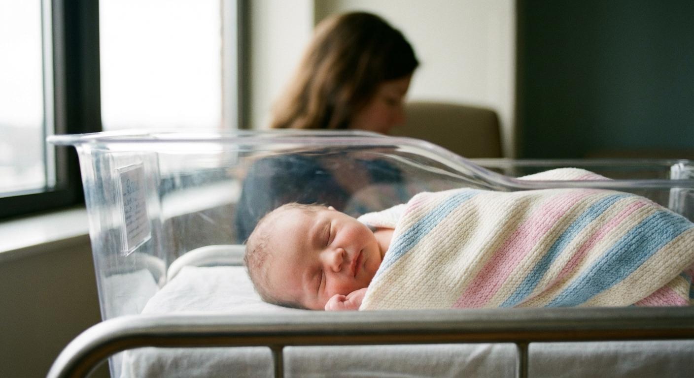
When a newborn looks yellow under hospital lights, parents may hear two very different sentences: “It’s common” and “We need to check quickly.” That emotional swing is exactly where hemolytic disease of the newborn sits. It can look like ordinary jaundice, yet the mechanism is immune-related, and bilirubin can rise fast.
The aim is straightforward: identify risk during pregnancy, recognise early signs after birth, treat jaundice and anaemia promptly, and plan follow-up so your baby remains safe and comfortable.
What hemolytic disease of the newborn means (HDN/HDFN)
Hemolytic disease of the newborn (also called HDN or HDFN) happens when a mother’s immune system makes IgG antibodies against red blood cell antigens on the baby’s blood cells. Antigens are tiny “identity markers” on the surface of red blood cells, often inherited from the father.
Those IgG antibodies can cross the placenta. Once they reach the baby’s circulation, they attach to the baby’s red blood cells and speed up their breakdown. This is haemolysis. No one “causes” this by eating the wrong food, missing a supplement, or lifting something heavy. It is an immune mismatch related to blood groups.

Doctors mainly keep an eye on two outcomes:
- Anaemia (low red blood cells, so oxygen delivery reduces)
- Hyperbilirubinaemia (high bilirubin), leading to jaundice (yellow skin and eyes)
Some babies only need a short spell of phototherapy. Others need closer monitoring, and a smaller group may need IVIG or transfusion-based care.
Names you may still hear
“Erythroblastosis fetalis” is an older term. It describes the baby’s body pushing out immature red blood cells (erythroblasts) because it is trying to compensate for ongoing haemolysis. Many teams now simply say hemolytic disease of the newborn.
IgG versus IgM: why placental crossing matters
A key detail is the antibody class:
- IgG crosses the placenta and can affect the fetus or newborn.
- IgM usually stays in the mother’s bloodstream and does not cross the placenta in the same way.
So when clinicians talk about hemolytic disease of the newborn, IgG is usually the driver.
Why doctors take it seriously: bilirubin, the brain, and severe anaemia
When red blood cells break down, they form unconjugated (indirect) bilirubin. Newborn livers are still maturing, so bilirubin processing and elimination are less efficient in the first days.
That is why clinicians focus on preventing:
- Significant anaemia (the heart may work harder to supply oxygen)
- Very high or rapidly rising bilirubin (bilirubin can irritate or injure the brain if it climbs too high)
- Hydrops fetalis (fluid build-up before birth, linked to severe fetal anaemia)
- Acute bilirubin encephalopathy and, rarely with timely care, kernicterus
You may be thinking: jaundice is so common, how do we know when it is different? Timing gives clues.
Hemolytic disease of the newborn versus physiologic jaundice
Many babies have mild jaundice. In general:
- Physiologic jaundice tends to appear after 24 hours and rises gradually, without anaemia.
- In hemolytic disease of the newborn, jaundice can appear within the first 24 hours (sometimes very early) and bilirubin may rise quickly.
- A direct antiglobulin test (DAT / direct Coombs) may be positive, showing antibodies attached to the baby’s red blood cells.
These patterns guide urgency, but they do not replace a clinician’s assessment.
How hemolytic disease of the newborn happens inside the body
Alloimmunisation: when antibodies target the baby’s red blood cells
Most hemolytic disease of the newborn is alloimmune: the mother lacks a particular antigen, the baby has it, and the immune system recognises it as “foreign” after exposure.
Placental transfer and extravascular haemolysis
IgG crosses the placenta via normal transport. In the baby, antibody-coated red blood cells are removed mainly in the spleen, called extravascular haemolysis.
What follows: anaemia and rising bilirubin
As haemolysis continues:
- Haemoglobin drops, leading to anaemia.
- The body compensates, leading to increased reticulocytes (young red blood cells), and sometimes enlarged liver and spleen (hepatosplenomegaly).
- Bilirubin production rises, increasing jaundice risk.
Severe end: hydrops fetalis and bilirubin-related neurologic risk
If fetal anaemia becomes profound, the heart can struggle and hydrops fetalis may develop (fluid in abdomen, around lungs or heart, and swelling under the skin). After birth, if unconjugated bilirubin becomes dangerously high, it can cross into brain tissue and cause bilirubin-induced neurologic dysfunction.
Causes and types of hemolytic disease of the newborn
Rh incompatibility (Rh disease)
Rh(D) incompatibility is the classic cause of severe disease when prevention is missed:
- An Rh-negative mother is exposed to Rh-positive fetal red cells (delivery is a common moment, but bleeding, procedures, or trauma can do it too).
- The immune system forms anti-D IgG.
- In a later pregnancy with an Rh-positive fetus, anti-D crosses the placenta and can trigger hemolytic disease of the newborn.
A practical point: the first pregnancy may be less affected if sensitisation happens near delivery, later pregnancies can be more affected because immune memory produces IgG faster.
ABO incompatibility (ABO disease)
ABO-related hemolytic disease of the newborn is common and often milder. It often occurs when the mother is blood group O and the baby is A, B, or AB, because some group O mothers have IgG anti-A and anti-B that can cross the placenta.
Typical pattern:
- Early jaundice is the main issue
- Anaemia is mild or absent
- DAT is often positive
- Phototherapy usually works well
It can happen even in a first pregnancy. Hydrops is rare.
Other antibodies (Kell, Duffy, Kidd, MNS)
Not all problems are Rh(D) or ABO.
- Anti-Kell can be severe because it may also suppress fetal red blood cell production (fetal erythropoiesis).
- Duffy and Kidd antibodies show variable severity.
- Some MNS antibodies can also cause clinically important disease.
Who may be at higher risk?
Risk increases with prior exposure to the antigen, such as:
- A prior pregnancy
- A prior blood transfusion
- In Rh-negative mothers, missed or delayed Rh immunoglobulin prophylaxis (anti-D prevention)
Certain events increase fetomaternal haemorrhage (mixing of fetal blood into maternal circulation):
- Bleeding in pregnancy (including placental abruption)
- Abdominal trauma
- Chorionic villus sampling or amniocentesis
- Some obstetric procedures
If the mother is Rh-negative and not sensitised, clinicians may advise Rh immunoglobulin after such events as per local protocol.
Prenatal screening and fetal monitoring
Early pregnancy labs: ABO/Rh typing and antibody screen
Routine antenatal bloodwork includes:
- ABO/Rh type
- Antibody screen (indirect Coombs)
If positive, the lab identifies the antibody, which shapes monitoring.
Antibody titres and “critical” thresholds
Many teams follow titres over time. A rising titre or reaching a lab’s threshold can trigger closer fetal surveillance. With anti-Kell, titres may not predict severity well, so monitoring may be more intensive even at modest titres.
Partner testing and fetal antigen testing
Depending on availability, clinicians may test the father’s antigen status. In some centres, fetal antigen status can be assessed with cell-free fetal DNA from maternal blood.
Ultrasound and MCA Doppler
Ultrasound can suggest anaemia or hydrops: ascites, effusions, skin swelling, enlarged heart, thickened placenta.
A key screening tool is middle cerebral artery Doppler (MCA-PSV). In fetal anaemia, blood flow velocity rises. Values around or above 1.5 MoM often suggest moderate to severe anaemia.
Prenatal management and fetal therapy
Prevention of anti-D disease with Rh immunoglobulin (RhIg)
For an unsensitised Rh-negative mother, RhIg given at recommended times prevents anti-D antibody formation. It is prevention, not treatment for existing antibodies.
Monitoring an alloimmunised pregnancy
When antibodies are already present, care focuses on:
- Following antibodies (often titres)
- Serial ultrasound and MCA Dopplers (timing depends on antibody type and prior history)
Intrauterine transfusion
If fetal anaemia is significant, intrauterine transfusion can raise fetal haemoglobin and may stabilise hydrops. This is performed in experienced centres with specially prepared blood.
Planning delivery
Delivery planning may include a hospital with NICU support and a blood bank ready with compatible, antigen-negative blood products, because some babies need intensive phototherapy, IVIG, or transfusion soon after birth.
What parents may notice: before and after birth
During pregnancy
Most warning signs are picked up on scans and Dopplers rather than symptoms you can feel.
In the first days after birth
Parents may notice:
- Yellowing of skin and eyes (especially early)
- Unusual paleness
- Fast breathing, sleepiness, tiring at feeds
- Feeds becoming shorter or less effective
If jaundice seems to spread down towards the legs, or your baby becomes less alert, seek medical review promptly.
Signs that need urgent assessment
Seek urgent care if jaundice is present with very poor feeding, marked sleepiness, breathing difficulty, widespread swelling, a high-pitched cry, abnormal movements, or seizures.
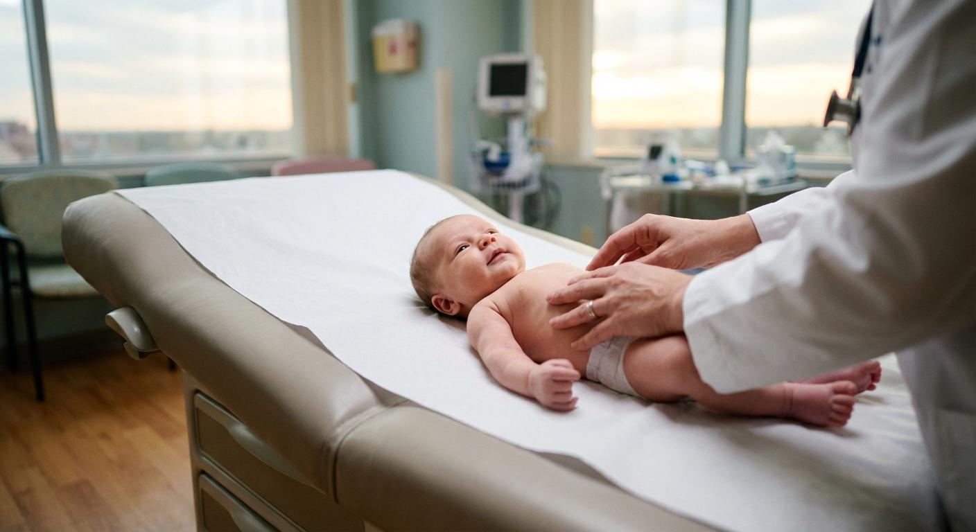
Postnatal diagnosis: tests used in hospital
To evaluate hemolytic disease of the newborn, clinicians may check:
- Baby’s blood group and DAT (direct Coombs)
- Haemoglobin/haematocrit
- Reticulocyte count
- Blood smear
- Serial bilirubin levels (rate of rise matters)
If DAT is negative, doctors may also consider other causes: G6PD deficiency (more common in some populations), hereditary spherocytosis, pyruvate kinase deficiency, infection, bleeding, or non-haemolytic jaundice.
Treatment after birth
The goals: protect the brain and correct anaemia
Treatment depends on age in hours, gestational age, bilirubin trend, and clinical signs.
Phototherapy
Phototherapy uses blue light to convert bilirubin into water-soluble forms.
During treatment, teams may monitor:
- Bilirubin frequently (sometimes every 4 to 6 hours initially in haemolysis)
- Temperature and hydration
- Weight, urine and stool output
- Feeding support
- Eye protection during lights
IVIG (in selected cases)
In some immune haemolysis cases, IVIG may be considered to reduce haemolysis and lower exchange transfusion need. Practice varies between units.
Red blood cell transfusion
If anaemia is significant, especially with symptoms like persistent fast heart rate, poor feeding, or breathing trouble, packed red blood cell transfusion may be advised, using newborn-appropriate compatible blood.
Exchange transfusion
Exchange transfusion may be used when bilirubin remains too high despite intensive phototherapy (and sometimes IVIG), or when severe anaemia is causing clinical impact. It rapidly lowers bilirubin, removes some antibodies, and replaces damaged red blood cells.
Going home and follow-up
Rebound jaundice
Because haemolysis can continue, a bilirubin recheck may be planned after phototherapy stops, often within 24 to 48 hours.
Late-onset anaemia
Maternal IgG can remain in the baby’s circulation for weeks, so late anaemia can appear after discharge. Follow-up blood tests may be scheduled.
Feeding and hydration
Frequent feeding helps hydration and stooling, which supports bilirubin removal. If feeding feels like hard work, ask early for lactation or paediatric support so intake is protected while your feeding goals are respected.
When to seek urgent care after discharge
Seek urgent medical care for worsening yellowing, very poor feeding, fewer wet nappies, fever (38°C/100.4°F or higher), breathing difficulty, unusual limpness or stiffness, abnormal cry, or abnormal movements.
Prevention and planning future pregnancies
For Rh-negative mothers who are not sensitised, timely RhIg during pregnancy and after delivery (if the baby is Rh-positive) prevents most anti-D related hemolytic disease of the newborn.
After sensitising events (bleeding, pregnancy loss, ectopic pregnancy, procedures, abdominal trauma), RhIg may be advised as per local protocol if the mother is Rh-negative and not already sensitised.
If a prior baby was affected, ask early for a plan: which antibody is involved, whether the fetus could carry the antigen, when MCA Dopplers should start, and where delivery should be planned.
To remember
- Hemolytic disease of the newborn is caused by maternal IgG antibodies crossing the placenta and breaking down the baby’s red blood cells.
- The main concerns are anaemia and rapidly rising bilirubin, early jaundice (within 24 hours) is a common clue.
- Common causes include Rh(D) and ABO incompatibility, but Kell and other antibodies can also be responsible.
- Treatment may involve phototherapy, IVIG in selected cases, transfusion, or exchange transfusion.
- There are professionals to support you. Your obstetrician, neonatologist, and paediatrician can explain results and follow-up clearly.
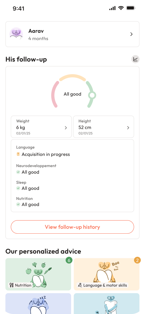
- You can also download the Heloa app for personalised guidance and free child health questionnaires.
Further reading :