Abdominal pain in children, worries about liver health, or sudden vomiting that refuses to stop—the list of parental anxieties around a child’s belly is long, and each new symptom has its own shadow of uncertainty. When doctors mention an “abdominal ultrasound,” do floodgates of questions open in your mind? Is it safe? Will my child feel any discomfort? Will this help us avoid other, scarier procedures? In today’s medical landscape, abdominal ultrasound has evolved into a trusted, gentle cornerstone for exploring what’s going on beneath the surface—without needles, without ionising radiation, and often without even needing to change into a hospital gown.
Let us walk together through the essentials: how this scan works, when it shines brightest, how to prepare your child (and yourself), the boundaries of what it can reveal, and how to decode the report that lands in your inbox a day or two later. Offers will be made for simple tips—how to keep things smooth in the waiting room, what questions to have in your mind, how to keep your child comfortable, and when to relax, reassured that no major mystery remains undetected.
What is an abdominal ultrasound? A glance at technology and technique
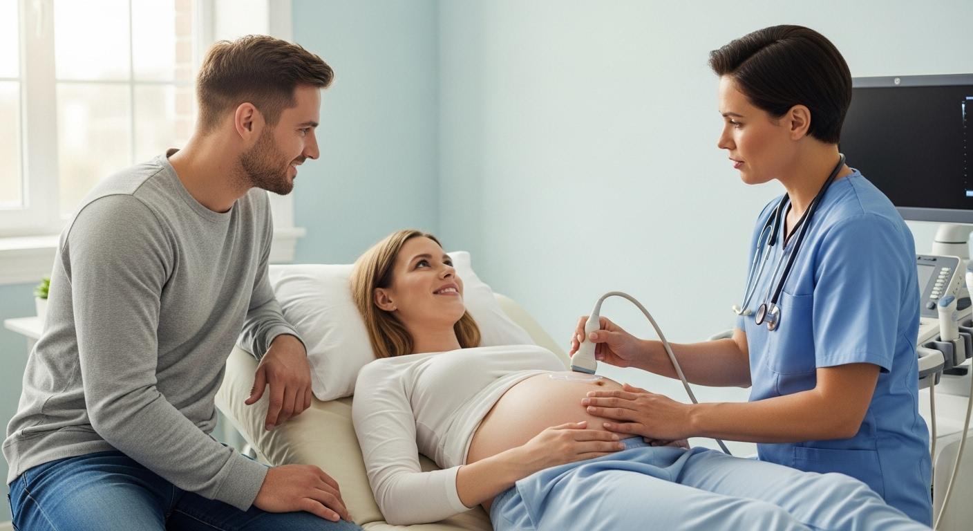
Imagine a scene: a quiet room, dimly lit; a technician rubs a slightly cool, water-based gel onto your child’s belly. A transducer—a small, wand-like device—glides gently over the skin. Inside, sound waves (completely harmless, cannot be felt or heard) travel invisibly through the tissues. These bounce back, creating an instant, moving portrait of the organs hidden beneath the abdominal wall. This is the promise of abdominal ultrasound: noninvasive and child-friendly.
Why is ultrasound such a favourite tool, especially in children and pregnancy?

- Immediate, real-time images capture the liver, gallbladder, bile ducts, pancreas, spleen, kidneys, urinary bladder, large blood vessels (like the abdominal aorta), and surrounding spaces.
- Particularly clever is the use of “Doppler mode.” With Doppler ultrasound, the movement of blood can be visualised, including flow in fragile vessels—helpful for picking up blockages or checking vessel health.
Medical vocabulary, reimagined: “Hyperechoic”—meaning bright or white on the scan, suggesting denser tissue; “Anechoic”—dark, indicating fluid. The science is sound, the safety record reassuring: abdominal ultrasound uses no X-rays, no radiation, and brings none of the long-term risks associated with CT scans.
Yet, even the best technology has its limitations. Air trapped in bowel loops sometimes “shields” the deeper structures, while a thicker layer of fat can lead to fuzzier details. Younger children may find it tough to stay still, affecting image clarity, but even here, paediatric teams bring experience and patience.
When is an abdominal ultrasound needed? Common triggers for parents
Emergencies and urgent concerns
Is your child doubled over in pain, vomiting persistently, or showing signs of dehydration? Medical teams listen carefully for these “red flags.” Some of the most pressing situations calling for an abdominal ultrasound include:
- Suspected appendicitis (right lower belly pain, fever, loss of appetite)
- Intussusception—when bowel telescopes into itself, seen as a “target sign” on the scan and often treated without surgery
- Hypertrophic pyloric stenosis in infants—persistent, forceful vomiting and visible muscle thickening on the scan
Other urgent uses:
- Sudden trauma: the “FAST” (Focused Assessment with Sonography in Trauma) scan checks for internal bleeding or collections
- Unexplained jaundice, concern for gallstones, or urine suddenly tinged red—here, an ultrasound can show gallstones, dilated bile ducts, or kidney stones
Timing is key. If there are signs of internal bleeding, or doctors suspect intussusception, the abdominal ultrasound may be done within minutes of arrival.
Chronic complaints and ongoing follow-up
Many health concerns evolve quietly, sometimes over months:
- Chronic liver disease: scanning for signs of fatty liver, cirrhosis, or abnormal blood flow in the portal vein
- Recurrent urinary tract infections in children: checking kidney size, structure, and looking for hydronephrosis (swelling, usually from blockage or reflux)
- Monitoring for abdominal masses detected on exam: A cystic lesion may look reassuring, while solid masses may need MRI for further clarity
Special mention: Screening for abdominal aortic aneurysm in certain adults (older age, long-term high blood pressure, or a smoking history).
What can be detected by abdominal ultrasound? A detailed tour
- Liver: Looks at contour, brightness, presence of focal lesions or steatosis (fatty infiltration), and examines the portal vein using Doppler for unusual flow patterns.
- Gallbladder and bile ducts: Finds gallstones, detects sludge (thick bile), IDs a thickened wall or tender points, and looks for dilated bile ducts suggesting an obstruction.
- Pancreas: May be partially hidden by bowel gas, but pancreatitis (inflammation, swelling, irregular borders) is often recognizable.
- Kidneys and urinary tract: Checks size, echo pattern of the cortex, presence of cysts, stones (usually casting a bright shadow), and hydronephrosis.
- Spleen: Evaluates enlargement, cysts, or signs of infection.
- Aorta and large vessels: Measures diameter, screens for aneurysms, and looks for plaques or thrombus.
- Bowel: Spots classic signs of appendicitis, intussusception, wall thickening, free peritoneal fluid, or localized abscesses.
A note for curious parents: Sometimes, if abnormalities are detected, a further scan using MRI or CT is suggested for more granularity and to guide treatment.
Types of abdominal ultrasound: Complete, targeted, and specialised
- Complete abdominal ultrasound: Covers all major organs within the abdomen, ideal for diffuse symptoms or unclear complaints.
- Targeted exams (“right upper quadrant,” “renal only,” etc.): Focused checks when doctors have already zeroed in on a likely culprit.
- Point-of-care ultrasound (POCUS) and FAST: Rapid bedside scans, used in emergency departments to answer urgent questions.
- Doppler studies: Looks at blood movement, especially valuable for checking vessel blockages or abnormal velocities.
- Contrast-enhanced ultrasound (CEUS) and elastography: Advanced tools for more precise tissue characterisation, particularly in the liver.
Special paediatric and pregnancy protocols exist. The choice of probe and technique varies—smaller probes and higher frequencies for children, adjusted for their size and physiology.
Preparation: Steps to make the day go smoothly
- Fasting: Often 6–8 hours before the scan. Skipping heavy, fatty meals and fizzy drinks can help reduce gas, improving clarity.
- Full bladder: For some pelvic or lower abdominal scans, especially in older children or adults, drinking 500–750 mL of water about an hour before the scan and holding urine is requested. This moves the bladder up, creating a better “window.”
- Medications: Do not stop your child’s regular medicines unless told specifically by your doctor. For diabetes or small children, ask about how to handle fasting—sometimes a small snack is allowed, or adjustments are made.
- Practical tips: Bring previous reports, referral letters, favourite toys (for distraction), and arrive a bit early. If fasting is missed and the scan is urgent, let the clinic know—they may still proceed or adapt instructions.
During the scan: What to expect, and comfort measures
- No pain, just mild discomfort: A little cold gel, some gentle pressure, maybe a ticklish feeling. The technician may ask your child to roll, take deep breaths, or hold still for a few seconds.
- Seated with you: Most centres welcome a parent in the room to offer reassurance, sing a song, or bring out a comforting storybook.
- How long?: Most abdominal ultrasounds take between 15 and 45 minutes, depending on what needs to be checked. Rest assured, no lasting discomfort lingers after the gel is wiped away.
Results, reporting, and the medical language decoded
- The report arrives. What might you see? Indication (the reason for the test), the organs and vessels examined, and a conclusion—often filled with terms like “no focal lesion,” “biliary dilatation,” or “hydronephrosis absent.”
- Follow-up explained: If a cyst or suspicious lesion is noted, MRI or specialised tests may follow. Sometimes, blood or urine tests are recommended to correlate findings.
- Next steps: Don’t hesitate to seek clarification on medical terms. Radiology teams are ready to explain images and suggest what comes next.
Limits and when additional imaging might be necessary
Abdominal ultrasound is powerful, but not infallible. Intestinal gas can block views, small tumours can hide, and overweight can blur fine detail. If needed, doctors may suggest repeating the scan, switching to MRI for more detail (attractively, no radiation here either), or moving to CT for rapid answers in emergencies.
Comparing abdominal ultrasound with other imaging choices
When does ultrasound stand tall, and when do other techniques step in?
- Ultrasound: Perfect for soft tissues, paediatrics, gallstones, and first evaluation of abdominal pain. Leaves no “radiation footprint.”
- CT scan: Speedy, detailed, especially if trauma, bleeding, or unusual calcifications are suspected. Uses ionising radiation.
- MRI: Unmatched for characterising liver and pancreas texture, or pinpointing soft tissue changes—drawback is duration and, sometimes, limited access.
- Scintigraphy: Rare, used for very specific conditions like organ function or unusual blood flow studies.
The usual progression: try abdominal ultrasound first. If clarity is lacking or more surgical planning is needed, advanced imaging comes next.
Practical, parent-centric tips for before, during, and after
- Before: Collect paperwork (referrals, previous tests), check instructions for fasting or full bladder, and schedule as early as possible (morning slots reduce hunger-triggered tears).
- During: Keep your tone light. Try a game or deep-breathing for reluctant children. It’s OK to ask for a break or pause.
- After: Life resumes as usual. Wait for formal report, and follow up promptly on advice or next steps if other tests are needed.
- Choosing the centre: Seek clinics with experience in paediatric scans, modern machines, and a willingness to involve parents.
Cost varies and may depend on preparation, urgency, and type of examination. Insurance often covers scans prescribed by a doctor; private-pay clinics may offer lower prices but fewer paediatric comforts.
Clinical snapshots
- Acute gallbladder pain with fever: Abdominal ultrasound may show small, moving gallstones, a thickened wall, and rim of fluid—suggesting inflammation.
- Monitoring fatty liver: A brighter, homogenous liver, with regular outline, may signal mild steatosis. Pulsed follow-up, nutritional guidance, and blood tests are part of the strategy.
- Aortic screening in older adults: Measurement above normal suggests an aneurysm—further checks or tight monitoring may be warranted.
Key Takeaways
- Abdominal ultrasound offers a gentle, effective, and noninvasive method to explore sudden or ongoing stomach complaints in both children and adults.
- Preparation makes a world of difference: fasting when asked, a full bladder when required, and avoiding fizzy drinks can spare repeat trips.
- Liver, gallbladder, kidneys, pancreas, spleen, blood vessels, and bowel—all can be evaluated, though some “blind spots” remain if gas or tissue blocks the view.
- If further detail is necessary, MRI is often the next radiation-free option; CT scans leap in for rapid or complex cases.
- Reports use medical terms—always seek explanations if doubts linger.
- Support is available. Reach out for help in understanding results, ask your healthcare team for clear plans, and advocate for your child’s comfort and wellbeing.
- For tailored advice, digital health tools like the application Heloa can provide free health questionnaires and personalised recommendations for your child’s care journey.
Questions Parents Ask
How reliable is abdominal ultrasound?
Abdominal ultrasound is highly informative in many situations, yet its reliability depends on the organ, the presence of air or fat, child’s cooperation, and clarity of the clinical question. Classic signs—like intussusception, gallstones, or hydronephrosis—are usually unmistakable. Smaller lesions, or structures deep beneath gas-filled bowel, might be missed. If there’s any doubt, your healthcare team may advise repeating the scan or opting for MRI or CT for more definitive evaluation.
How quickly will results be available?
Usually, the technician or doctor might give you a brief verbal impression just after the scan, especially if things look normal or there’s an urgent concern. The full radiology report can appear within 24–48 hours for elective scans, and on the same day for emergencies. Many centres update status online, but if waiting feels anxious, don’t hesitate to check with the front desk or request a timeline.
How much does an abdominal ultrasound cost, and is insurance involved?
Expect variability—costs differ by healthcare setting, scan type (simple, Doppler, or contrast-assisted), and urgency. When a doctor prescribes the scan, insurance often provides full or partial coverage. In private clinics or independent centres, charges can differ. If budget poses a concern, inquire at reception or with your insurer for an estimate, explore alternate centres, or ask about instalment plans. Openness about financial worries is always encouraged—many facilities assist with practical solutions.

Further reading :