You want to keep your child safe, comfortable, and well cared for. When a doctor mentions an abdominal ultrasound, you may wonder whether it hurts, how accurate it is, or whether it is really necessary. Those are fair questions. Here is a clear path forward, from what the scan shows to how to prepare, with practical steps to ease the day and confident language you can use when talking with your care team.
What an abdominal ultrasound is and why it helps
An abdominal ultrasound is a real time scan that uses sound waves to create pictures of the organs in the belly. No X rays, no sedation for routine exams, and no needles to begin. A small handheld probe slides over warm gel on the skin and sends sound waves into the body, then listens to the echoes. The machine turns those echoes into moving images.
You might ask, what does that look like in practice. A sonographer adjusts settings to balance depth and detail. High frequency probes give crisp surface detail, low frequency probes reach deeper structures like the liver or aorta. When vessels matter, color and spectral flow tools, for example Doppler, show direction and speed of blood flow.
Abdominal ultrasound is often first line in children and during pregnancy. Why. It is fast, portable, and radiation free. The scan adapts to breathing and positioning, which is helpful for little bodies and expectant parents.


- Typical organs reviewed, liver, gallbladder, bile ducts, pancreas, spleen, kidneys, bladder, major vessels like the aorta and vena cava
- Optional flow assessment, such as portal vein flow, renal artery velocities, and aortic waveform patterns
- Real time observation, movement of bowel loops, sliding of tissues, and fluid shifts during gentle pressure
Safety is a common question. Ultrasound uses non ionizing radiation, which means energy levels that do not break chemical bonds. Teams also follow the ALARA principle, as low as reasonably achievable, which keeps exposure time and power settings at sensible minimums while still answering the clinical question.
When to consider imaging
Urgent symptoms that merit rapid abdominal ultrasound
If your child has severe or worsening belly pain, repeated vomiting, fever with abdominal pain, signs of dehydration, or looks very unwell, imaging may be needed the same day. Ultrasound can rapidly clarify several high stakes conditions without radiation.
- Suspected appendicitis, appendicitis ultrasound looks for a non compressible, enlarged appendix, surrounding inflammation, and sometimes an appendicolith
- Intussusception, classic bullseye or target sign, the scan can both diagnose and help guide a non surgical reduction in many centers
- Projectile vomiting in a young infant, suggestive of hypertrophic pyloric stenosis, ultrasound measures pyloric muscle thickness and channel length
- Trauma, a focused bedside exam often called point of care ultrasound screens for internal fluid that may reflect bleeding
- Right upper quadrant pain or jaundice, ultrasound checks for gallstones, sludge, wall thickening, or dilated bile ducts
- Renal colic, scans look for kidney stones and upstream dilation of the collecting system called hydronephrosis
Timing depends on severity. Intussusception or signs of internal bleeding are immediate concerns. Appendicitis and concerning jaundice usually warrant same day evaluation.
Ongoing issues, follow up, and screening
For persistent or recurrent concerns, abdominal ultrasound can track change over time and steer next steps.
- Liver, fatty change called steatosis appears brighter, cirrhosis reshapes contours and can enlarge the spleen, focal nodules are described and may lead to MRI for more detail
- Kidneys and urinary tract, ultrasound evaluates size, cortex, cysts, stones, and degrees of hydronephrosis, it also helps in recurrent UTIs and congenital variants
- Aorta and large vessels, noninvasive measurement of aortic diameter helps screen for aneurysm risk and checks for mural thrombus
- Oncology follow up, known lesions or postoperative sites can be monitored, with MRI or CT added if needed
- Pediatric recovery checks, after pyloric surgery, intussusception reduction, or appendicitis with abscess, ultrasound confirms resolution without radiation
What an abdominal ultrasound can show, organ by organ
Liver
The liver is mapped for size, texture, and any focal lesions. A fatty liver often looks brighter and more uniform, doctors may call that hyperechoic. Cirrhosis can give an irregular outline and may be paired with a larger spleen. Simple cysts are fluid filled and look anechoic, meaning black without internal echoes. Flow analysis examines the portal vein and hepatic artery if indicated.
Gallbladder and bile ducts
Gallstones are dense and cast a clean shadow behind them. Wall thickening plus tenderness with probe pressure raises concern for inflammation. Dilated bile ducts suggest blockage in the common channel.
Pancreas
This gland can be tricky to see because bowel gas blocks some views. Acute inflammation makes it puffy and more echo rich or sometimes mixed in appearance. Small deep lesions sometimes require MRI or CT if concern remains after abdominal ultrasound.
Kidneys, ureters, and bladder
Size, shape, and cortex are recorded. Stones are bright with a posterior shadow. Dilation of the collecting system, called hydronephrosis, is graded by severity. Some children have duplicated collecting systems or ureteroceles, both visible on ultrasound.
Spleen
Usually homogeneous, the spleen is measured and checked for cysts, infarcts, or abscesses. Enlargement, called splenomegaly, can be linked with infections or blood conditions.
Aorta and major vessels
The aorta is measured outer to outer in millimeters to screen for enlargement. Flow velocity patterns on spectral analysis can suggest narrowing.
Bowel and fluid
With graded pressure, also called graded compression, the sonographer can evaluate the appendix and right lower quadrant. Intussusception shows the classic target sign. Bowel wall thickening and free fluid raise the index of suspicion for inflammation or obstruction.
Types of abdominal ultrasound and how they are used
- Complete exam, a broad survey when symptoms are not localized
- Targeted exam, focused questions like right upper quadrant or renal only or aortic check
- Bedside or emergency uses, point of care ultrasound and trauma fast exam for quick decisions
- Flow assessment, color and spectral tools, power based modes such as power Doppler
- Specialized options, contrast enhanced ultrasound improves characterization of some lesions, and elastography estimates tissue stiffness such as liver fibrosis
Probe selection matters. A belly survey commonly uses a curvilinear probe for depth, and a linear probe for high detail in children or superficial structures.
How to prepare, practical guidance for families
Preparation boosts image quality and lowers the chance of a repeat visit. You are not being picky by asking for clear instructions, you are being wise.
- Fasting, often 6 to 8 hours for older children and adults, 4 to 6 hours for many children, this empties the gallbladder and reduces bowel gas
- Food tips, skip high fat meals and fizzy drinks the day before and the day of the scan, avoid chewing gum which increases swallowed air
- Hydration, usually encouraged for renal studies unless medically restricted
- Bladder strategy, some abdominopelvic views need a partially full bladder, older kids and adults often drink 500 to 750 milliliters an hour before the scan without voiding
- Medication, continue essential medicines unless your clinician advises otherwise, for diabetes, discuss fasting and bring glucose supplies and a snack for after the scan
- Comfort items, a favorite small toy or book can help a child settle, a parent by the bedside calms the room
Why fasting. The gallbladder shrinks when you eat, which hides stones and wall detail. Why a full bladder. Fluid acts as an acoustic window to improve the view of pelvic structures.
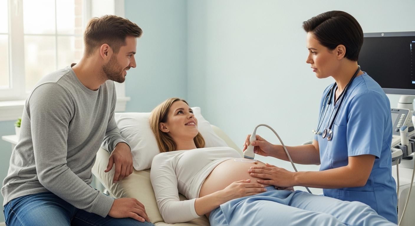
What happens during the exam, step by step
Arrival is straightforward. You check in, confirm preparation, and may change into a gown. The sonographer explains the plan in simple terms.
- Your child or you lie on the table
- Warm gel is applied to the skin
- The probe glides with gentle pressure, pausing for pictures and short clips
- You may be asked to take a deep breath or hold it briefly, this moves organs into a better window
- Flow tools are added when needed for vessels like the portal vein or renal arteries
How long. Most abdominal ultrasound sessions take 15 to 45 minutes, depending on the question and how easily clear images are obtained. The gel may feel cool, the scan itself should not hurt.
Limits, safety, and when another test is needed
Every test has limits. Bowel gas can block the view, body habitus and dressings can reduce detail, and tiny lesions can fall below the resolution of sound waves. There are almost no absolute reasons to avoid abdominal ultrasound in children or during pregnancy. Gel allergy is rare, alternatives exist.
When images are not clear enough, your team may suggest a repeat exam, MRI for detailed liver or pancreas assessment, or CT when speed and a full overview are essential. Blood and urine tests support imaging by adding biochemical context.
Understanding the report and common phrases you might see
A standard report organizes the story in a consistent way. Indication, what was examined, techniques used, measurements, organ descriptions, and a final summary called the impression.
- Normal study, organs of expected size and appearance, no duct dilation, no free fluid
- Gallstones, stones with wall thickening and focal tenderness, matched to symptoms and labs
- Simple liver cyst, a well defined round fluid space without internal flow
- Indeterminate lesion, a heterogeneous mass that needs MRI for full characterization, sometimes a needle sample is discussed if safe and appropriate
Words to know. Echogenic or hyperechoic means brighter, hypoechoic means darker, anechoic means purely black fluid, and complex often means a mix of fluid and tissue.
Abdominal ultrasound compared with other imaging options
- Ultrasound strengths, no radiation, dynamic real time views, excellent for gallbladder disease, hydronephrosis, pediatric emergencies like appendicitis or intussusception, and vascular screening
- CT, very rapid and detailed, helpful for complications and calcifications, uses ionizing radiation
- MRI, outstanding soft tissue detail without radiation, longer exam and may be less available
A sensible path is to start with abdominal ultrasound when appropriate. If the answer is clear and matches the symptoms, move forward with care. If the answer is uncertain, MRI is usually preferred to avoid radiation, CT is chosen when speed or a complete overview will change decisions.
Special populations, what parents should know
Pediatrics and infants
Children benefit from ultrasound first strategies for appendicitis, intussusception, and pyloric stenosis. Age adjusted normal ranges guide interpretation, and probe choice matches body size. Positioning, distraction, and a calm room make a big difference.
Pregnancy, high BMI, and critical illness
In pregnancy, abdominal ultrasound and obstetric views are safe. A small tilt to one side can improve comfort and circulation. In higher body mass index settings, sonographers try multiple windows and specialized probes. In intensive care, fast bedside scans guide urgent choices.
Practical tips before, during, and after the scan
- Before, bring the referral, prior reports, and a medication list, confirm fasting and bladder instructions, morning times can make fasting easier for kids
- During, simple explanations and a quiet game reduce anxiety, ask for a brief pause if pressure feels uncomfortable
- After, normal activities resume right away, share the report with the prescribing clinician, schedule any complementary tests promptly if advised
Choosing where to go. Look for pediatric experience, modern equipment with flow tools, reasonable appointment times, and a unit that welcomes parent presence.
Frequently asked questions, quick clarity
- Does an abdominal ultrasound hurt, No, it should not, pressure may feel firm at times
- Can my child eat, Follow fasting instructions, small sips of water are sometimes allowed, confirm with the team
- Will my child be exposed to radiation, No, ultrasound uses sound waves, not X rays
- What if the appendix is not seen, The report may say nonvisualized appendix, doctors integrate symptoms, blood tests, and sometimes repeat imaging
- What about advanced tools, Contrast agents for ultrasound are microbubbles that enhance blood flow detail, they are used in selected cases and in specialized centers
Clinical snapshots to make it concrete
- Right upper quadrant pain with fever, abdominal ultrasound shows mobile stones, a thickened gallbladder wall, and a thin rim of fluid, initial medical care is started and surgery is considered if symptoms persist
- Fatty liver follow up, the liver appears bright and uniform without suspicious nodules, care focuses on nutrition, activity, and lab monitoring with a scheduled recheck
- Aortic screening in an older adult with risk factors, an enlarged diameter is measured, the plan includes surveillance or angiography based on size and growth rate
Key takeaways
- Abdominal ultrasound is safe, fast, and often first line for belly pain, vomiting, jaundice, and many pediatric scenarios
- Preparation matters, fasting when asked, skipping fizzy drinks and fatty meals, and having a full bladder when requested improves clarity
- The scan shows liver, gallbladder and ducts, pancreas, spleen, kidneys, bladder, vessels, and bowel, flow tools add hemodynamic insight
- Limits exist, gas, body habitus, and very small lesions can reduce visibility, when answers are uncertain, MRI or CT may be suggested
- Reports follow a clear structure with an impression and next steps, ask for plain language explanations if anything is unclear
- There are resources and professionals to support your decisions, for personalized tips and free child health questionnaires, you can download the application Heloa
Abdominal ultrasound gives answers without radiation, and with preparation and the right questions, it becomes a calm, informative step in your child’s care.
Questions Parents Ask
How accurate is an abdominal ultrasound?
Ultrasound is a very useful first test, but its accuracy depends on several factors: what organ is being looked at, the child’s size and how much bowel gas is present, how well the sonographer can visualize the target, and the specific question being asked. For some problems it is highly reliable — for example, the classic signs of intussusception are usually clear, and gallstones and hydronephrosis are commonly well seen. For others, especially very small lesions or deep structures blocked by gas (like parts of the pancreas), ultrasound may miss findings. If images are limited or the clinical picture remains uncertain, teams commonly recommend a repeat ultrasound or a more detailed exam such as MRI or CT to be sure. Rest assured, your care team will explain what the scan shows and suggest the next step if more clarity is needed.
How soon will we get the results?
You often get an immediate, informal update from the sonographer right after the scan — a quick reassurance or note about whether images were clear. The formal report interpreted by a radiologist may take longer: in routine outpatient settings it often arrives within 24–48 hours, while urgent or emergency exams are usually reported the same day. Many clinics also post results to a patient portal once finalized. If waiting is stressful, you can ask the clinic when to expect the official report and whether a clinician will review the findings with you by phone or at a follow-up visit.
How much does an abdominal ultrasound cost, and will insurance cover it?
Costs vary widely by country, facility, and the type of exam (a focused study, a complete abdominal survey, or a contrast-enhanced or Doppler study can change the price). In many health systems an ultrasound ordered by a clinician is covered or partly covered by insurance; in private-pay situations fees differ between hospital radiology departments and independent imaging centers. If cost is a concern, you can call the imaging center ahead and ask for an estimate, check with your insurer about coverage and preauthorization, or ask whether there are lower-cost community options. Don’t hesitate to mention financial worries — many centers can suggest payment plans or alternatives.

Further reading :