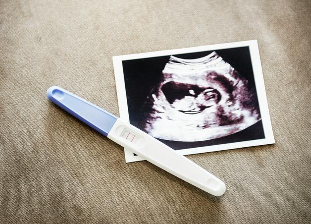
You see the term nuchal translucency in a report or hear it mentioned at a scan, and a dozen questions bubble up at once. Is this normal, what does the number mean, when do we worry, what are the next steps if it is higher than average. Let us set the stage clearly. Nuchal translucency is a measurement taken in early pregnancy that shifts probability, it does not label a baby, it guides smart choices. You will find the what, the when, the how, and most importantly, the so what, explained in plain language, with medical accuracy and practical routes forward. Ready to turn uncertainty into a plan you can live with.
Nuchal translucency, how it helps screen in early pregnancy
What the measurement is and why it matters
Nuchal translucency is the thin fluid layer behind a baby’s neck that can be seen during a first trimester scan. Some fluid is expected. A thicker layer can reflect differences in how lymph channels drain, how blood returns to the heart, or how connective tissue develops. That is why nuchal translucency acts as a screening marker for conditions such as Down syndrome and certain heart differences. It adjusts probability. It does not confirm a diagnosis.
Parents often ask, how much is too much. Think of nuchal translucency like a dimmer switch rather than an on off light. As the number rises, the chance of some conditions increases, and that is the cue to consider further testing or specialist imaging.
To keep terms consistent with research and clinical pathways, you will also see the technical tag nuchal_translucency used in guidelines and databases.


Difference between nuchal translucency, nuchal fold and cystic hygroma
These terms look similar, yet they describe different things and different timing.
- Nuchal translucency, a fluid space measured in the first trimester.
- Nuchal fold, a soft tissue thickness checked in the second trimester, it is not a fluid pocket, it is a fold of tissue.
- Cystic hygroma, a larger, often septated lymphatic finding that carries a higher chance of chromosomal or syndromic conditions, and may be linked with early swelling called hydrops.
You may also hear about the nuchal_fold, which is a second trimester marker and should not be confused with first trimester nuchal translucency.
When to have the nuchal translucency scan
Optimal timing and technical windows
Timing matters because the measurement is only reliable within a specific growth window. The ideal range is 11 weeks to 13 weeks and 6 days, when the crownrumplength or CRL is about 45 to 84 millimeters. Before 11 weeks the image is small and margins are harder to define, after 14 weeks the anatomy changes quickly and comparisons with reference charts become less dependable. Accurate dating, tied to gestational_age, is the baseline for any next step.
For clarity and standardization, programs often refer to the firsttrimesterscreen window when scheduling this visit.
Typical values and practical landmarks
At 11 to 12 weeks, the median nuchal translucency sits around 1.4 to 1.6 millimeters. Many programs consider 3.5 millimeters or more as clearly elevated, though interpretation is always adjusted for the baby’s size on the day. A simple mental map helps:
- Near 1.5 mm, very common, continue routine screening.
- Around 2.5 mm, mildly increased, consider noninvasive follow up like NIPT.
- At or above 3.5 mm, high enough to discuss deeper evaluation.
You may also see the measurement expressed as a multiple of the median, called MoM, that standardizes the value for the exact crownrumplength.
How the nuchal translucency scan is performed
Scan methods and what to expect
A typical appointment lasts 15 to 30 minutes. The sonographer starts with an abdominal scan, and if the view is limited, a transvaginal route can provide a clearer picture. Both are safe and painless. Expect coaching to help the baby rest in a neutral position, perhaps a short walk or a gentle wiggle to change the angle. If the ideal view is not captured, a brief rescan may be suggested.
In reports and training materials you may see technical terms like ultrasound, transabdominalultrasound, and transvaginalultrasound. These simply describe the scan route used to get the best view.
Image acquisition, measurement technique and quality assurance
A correct nuchal translucency requires a true midline side view, called a sagittalview, with clear visibility of the skin and the thin amniotic membrane. Calipers touch the inner borders of the translucent area at its thickest point. Several measurements are taken, the most reliable is recorded to the nearest tenth of a millimeter. High quality services save key images, review statistics, and follow clinicalguidelines with routine audits.
If an optimal plane cannot be obtained the first time, a repeat appointment or a referral to a fetal medicine specialist is reasonable and common.
Understanding nuchal translucency results and reporting
Millimeters, MoM and likelihood of conditions
Nuchal translucency is reported in millimeters and sometimes as MoM, which compares your baby’s measurement with the expected median at the same size. An MoM around 1 suggests a value near the expected center, higher MoMs signal a higher chance of certain conditions. Programs combine your baseline chance linked to maternalage with the nuchal translucency MoM to calculate pretestrisk and posttestrisk as part of a broader riskassessment.
Reports often present a number like 1 in 450 or 1 in 120. These figures help decide whether to offer a noninvasive screen or a diagnostic test.
Common thresholds and triage
To organize follow up, many teams use tiers.
- If the calculated risk is higher than about 1 in 250, invasive diagnostic testing is offered.
- If the risk falls between about 1 in 250 and 1 in 1000, noninvasive screening is offered.
- If risk is below about 1 in 1000, routine care continues, with optional additional screening.
Separately, an absolute nuchal translucency of 3.5 millimeters or more usually prompts a conversation about detailed imaging and diagnostic options, even if the calculated number sits near a boundary.
Combined first trimester screening, nuchal translucency plus blood tests
What tests are combined and why
The combined screen uses nuchal translucency together with maternal blood markers like PAPP A and free beta hCG, then integrates maternal age and pregnancy details. These markers shift in predictable patterns in pregnancies affected by common trisomies. This is often labeled the firsttrimestercombinedtest, and you may see blood markers grouped as maternalserummarkers such as freebeta_hCG.
Performance and limitations
Nuchal translucency alone detects about 70 to 75 percent of Down syndrome cases. Adding blood markers raises sensitivity to about 85 percent, with a false positive rate near 5 percent. Cell free DNA based screening, often called NIPT or cellfreeDNA_testing, performs even better for the common trisomies, but it remains a screen, it still needs confirmation if positive. No screening approach catches every condition, so false negatives and false positives do occur.
What an increased nuchal translucency can indicate
Chromosomal, genetic and structural causes
A higher nuchal translucency is associated with chromosomal conditions such as trisomy21 or downsyndrome, trisomy18 or Edwards syndrome, trisomy13 or Patau syndrome, and turnersyndrome. It is also linked with differences in the heart’s structure, which is why a focused fetal echocardiogram is often advised. These fall under the umbrella of fetalaneuploidy and structural conditions like congenitalheartdefects.
Sometimes the increased measurement relates to a cystic hygroma or early generalized swelling called hydrops. Single gene syndromes, such as Noonan spectrum, can also be part of the picture, especially with very high values or additional ultrasound clues. More rarely, infections like cytomegalovirus or parvovirus may contribute. And yes, sometimes every test returns reassuring and the pregnancy continues normally. That is why decisions move step by step.
Next steps after a high nuchal translucency, counseling and testing options
Noninvasive and diagnostic testing pathways
Think of your next step as a fork with two well marked paths. One path is noninvasive screening such as NIPT, which checks placental DNA fragments in the mother’s blood. If it suggests a high chance of a condition, the result needs confirmation through diagnostic_testing.
The other path is an invasive diagnostic test. Options include CVS or chorionicvillussampling, available from about 10 weeks, and amniocentesis, available from about 15 weeks. These can provide a karyotype, a chromosome level view of the baby’s genetic material, sometimes a microarray for smaller changes, and even sequencing when indicated. The risk of a procedure related loss is low, typically a few tenths of a percent. Timing, the level of certainty you want, and personal values guide the choice.
This is a good moment for prenatal_counseling, where benefits, boundaries, and timelines are discussed in a supportive setting.
Imaging and follow up
Detailed first trimester ultrasound, followed by targeted scans, refines what we know. A fetal echocardiogram is often scheduled when nuchal translucency is high. Follow up may include watching the nuchal area as the weeks pass, checking for signs of hydrops, and monitoring growth. If chromosome testing is normal but concerns remain, the team may suggest gene panels or exome sequencing, tailored to ultrasound patterns and family history. The result of this process can be a specific answer, or a continued plan of observation with clear checkpoints.
Nuchal translucency versus other screening strategies
NT compared with NIPT and second trimester serum tests
NIPT outperforms nuchal translucency for the three common trisomies, and many programs use it as a primary or contingent screen. Second trimester serum tests add coverage for conditions like neural tube defects. Yet nuchal translucency still matters. It provides an early look at anatomy, it can flag a higher chance of cardiac or syndromic conditions, and it helps decide who benefits from specialist imaging or diagnostic testing. Screening tools work best when they complement each other.
Role of NT in contemporary screening pathways
Even in an era of advanced DNA screening, nuchal translucency offers value for early structural assessment, integrated risk estimates, and triage for fetal echocardiography or genetic evaluation. Programs align these steps with published clinical_guidelines to keep care consistent and transparent.
Special situations and technical challenges
Twins, higher order multiples and ART pregnancies
In twins and higher order multiples, nuchal translucency and size are measured for each baby. Interpretation is more layered in monochorionic twins who share a placenta, so results are often reviewed with a fetal medicine specialist. In assisted reproduction, careful timing and chorionicity assessment are especially important. If images are limited by position or maternal factors, a short interval rescan or an alternative screening route may be suggested.
Technical and maternal factors affecting measurement
Maternal body weight, uterine tilt, fibroids, and the baby’s posture can influence image quality. Operator training, accreditation, and consistent technique improve reliability. If a value seems out of step with the image or the growth, ask for an image review or a repeat measurement. That is good practice, not a setback.
How risk is calculated and communicated
Risk is calculated in stages. A baseline chance tied to maternalage becomes a pretestrisk. The nuchal translucency MoM shifts that baseline using a likelihood ratio. Blood markers add another layer. The result is a posttestrisk that guides whether to consider cellfreeDNAtesting or move directly to a diagnostic option. Clear reporting includes the nuchal translucency in millimeters, the CRL, the MoM if available, and the final number with suggested next steps. When you read a risk like 1 in 300, an honest conversation about what that means for your family is part of the package.
Other first trimester ultrasound markers worth knowing
When seen with an increased nuchal translucency, some findings increase the chance of an underlying condition.
- A very small or absent nasal bone.
- Tricuspid regurgitation on Doppler.
- Early signs of hydrops, such as skin edema or fluid around organs.
- Size smaller than expected for dates.
- A small, transient umbilical herniation.
The more these cluster, the stronger the case for detailed genetic evaluation and careful anatomical follow up.
Accuracy, quality assurance and program standards
High performing services follow written protocols, use standardized planes, and convert nuchal translucency to MoM based on the crownrumplength. They review service wide statistics, store images, and maintain training to keep the median of MoMs near 1 with a stable spread. Consistent technique and accurate dating are what make results dependable over time.
Preparing for the appointment and reading your report
Practical checklist for parents
- Bring any dating information and prior scan reports.
- Ask whether blood markers will be drawn the same day, especially if requesting a combined screen.
- Wear comfortable clothing suitable for abdominal and possibly transvaginal imaging.
- Confirm coverage and costs in advance, note your questions about technique, accreditation, and follow up plans.
Understanding your nuchal translucency report
You will usually see the nuchal translucency in millimeters, the CRL, the dated gestational_age, and a risk estimate if calculated. The result may appear as a number like 1 in 300 or a category such as low, intermediate, high. Recommendations might include genetic counseling, NIPT, repeat imaging, fetal echocardiography, or a diagnostic test. If anything is unclear, ask your clinician to walk you through the numbers and options, step by step, in plain language. That conversation is part of good care.
Emotional support and shared decision making
Waiting is hard. Setting a simple plan helps. Write down the two or three things you need to know next, agree on a timeline for results, and choose small comforts while you wait. Ask questions that center your values. For example, what does this number mean for our baby, what are the timelines if we want a diagnostic answer, and given our priorities, what would you recommend and why. Thoughtful decisions come from clear information and steady support.
Helpful visuals and how they support understanding
Many parents find these visuals helpful at appointments.
- A sagittal profile diagram that shows exactly where nuchal translucency is measured.
- An annotated image that labels the scan route, gestational age, nuchal translucency value, and CRL.
- A graph of medians and reference limits that shows why MoM conversion matters.
- A simple flow from nuchal translucency measurement, to risk calculation, to offers of noninvasive screening or diagnostic testing, then to targeted imaging based on thresholds.
Sources and references to explore
- Fetal Medicine Foundation, NT measurement protocols and quality standards.
- Snijders et al., Lancet 1998, foundational multicenter data on nuchal translucency screening.
- Salomon et al., 2009, reference curves widely used in practice.
- Contemporary reviews on NIPT performance and the value of fetal echocardiography after increased nuchal translucency.
Key takeaways
- Nuchal translucency adjusts probability, it guides next steps, it does not diagnose on its own.
- The optimal window is 11 weeks to 13 weeks and 6 days, with accurate dating by crownrumplength for reliable interpretation.
- Millimeters and MoM tell the same story through different lenses, decisions hinge on the final personalized risk.
- A structured pathway exists, noninvasive screening, diagnostic testing when indicated, targeted imaging, and follow up that matches what is found.
- Programs that follow consistent clinical_guidelines and invest in quality assurance provide the most reliable results.
- There are many resources and professionals ready to support you, and you can download the Heloa app for personalized advice and free child health questionnaires at this link, https://app.adjust.com/1g586ft8.
Nuchal translucency can be a moment of uncertainty, yet with clear timing, solid technique, and well chosen next steps, it becomes a powerful tool for informed, calm decisions.
Questions Parents Ask
How long will I wait to get my nuchal translucency result and any follow-up tests?
You’ll often get the measured NT value at the end of the scan or soon after; a formal written report may take a few days. If blood markers are done at the same visit, combined-screen results typically arrive within several days to a week depending on the lab.
If you move to NIPT, most labs return results in about 5–14 business days (some offer faster options). For diagnostic testing: CVS can give rapid targeted results in a few days for common chromosomal changes, while a full karyotype or microarray may take one to three weeks. Fetal echocardiography or repeat specialist scans are usually arranged within days to a couple of weeks, depending on urgency and local access.
It’s completely reasonable to ask your care team for expected timelines and who to contact with questions — knowing the schedule often eases the waiting.
How much does a nuchal translucency scan cost and is it covered?
Costs vary widely by country, clinic and whether the scan is part of routine publicly funded care or done privately. In many public health systems an NT scan and medically indicated follow-up are covered; in private settings there can be out-of-pocket fees. Additional tests (NIPT, CVS, amniocentesis, microarray) have their own costs and coverage rules, and NIPT is often more expensive than the ultrasound.
Before your appointment, you can ask the provider for an estimated price list and check with your insurer about codes and coverage criteria. If finances are a concern, mention it — many clinics can explain cheaper options, payment plans, or when a test is likely to be covered for medical reasons.
How do you pronounce “nuchal translucency”?
Say it slowly like this: NOO-kul trans-LOO-sen-see.
Breaking it down helps: “nuchal” = NOO-kul, “translucency” = trans-LOO-sen-see. Don’t hesitate to ask the sonographer or clinician to say it again or to write it down — it’s perfectly okay to want the word pronounced clearly.

Further reading:
- Nuchal translucency — Knowledge Hub: https://www.genomicseducation.hee.nhs.uk/genotes/knowledge-hub/nuchal-translucency/
- First Trimester Screening, Nuchal Translucency and NIPT: https://www.hopkinsmedicine.org/health/treatment-tests-and-therapies/first-trimester-screening-nuchal-translucency-and-nipt
- Nuchal translucency measurement: https://www.mayoclinic.org/nuchal-translucency-measurement/img-20007028