By Heloa | 24 November 2025

Third trimester ultrasound essentials for confident birth planning

3 minutes
de lecture
Smiling mom-to-be on the exam table holding her partner's hand for her 3rd trimester ultrasound

By Heloa | 24 November 2025

Third trimester ultrasound essentials for confident birth planning

3 minutes
Smiling mom-to-be on the exam table holding her partner's hand for her 3rd trimester ultrasound

Par Heloa, le 24 November 2025

Third trimester ultrasound essentials for confident birth planning

3 minutes
de lecture
Smiling mom-to-be on the exam table holding her partner's hand for her 3rd trimester ultrasound

Feeling your baby’s kicks become stronger, noticing your belly grow a little more each week, perhaps lying awake at night as excitement and worries mix together — the third trimester is a journey like no other. Amidst this whirlwind, the third trimester ultrasound becomes a central moment. Maybe friends shared their experiences. Maybe you have questions lingering — Should you expect another scan? What exactly will it reveal when birth is just around the corner? If concerns about fetal growth, presentation, fluid levels, or the placenta keep cropping up, you are far from alone. Let’s untangle the medical facts, walk you through the process, highlight the decisions that may come up, and help you step into your next appointment ready and reassured.

Explore what makes the third trimester ultrasound distinct, how it checks your baby’s growth, detects position and health signs, measures amniotic fluid and placenta, and what insights it brings for your birth plan. Expect clarity on preparation, what the scan evaluates — and, in case an abnormality does appear, what happens next. Let’s journey together through evidence-based information, practical tips, and a rhythm that matches the pace of late pregnancy itself.

What Is a Third Trimester Ultrasound? Distinct Focus and Timing

A third trimester ultrasound is not just a repeat of the scans you might have had earlier in pregnancy. It arrives typically between 28 and 42 weeks — with a strong preference for the 31–33-week window (near 32 weeks for many). Unlike the extensive anatomy scan from the second trimester, this time the spotlight shifts. Now, clinicians are less focused on cataloguing every bone and organ shape, instead aiming to chart fetal growth, map out the amniotic fluid (AFI or deepest vertical pocket), track the placental location, verify presentation (head-down, breech, or other), and use focused Doppler studies where necessary to check for blood-flow patterns in vital vessels.

So, what does this mean in practice?

  • Is your baby growing as expected on the curve, or is there a hint of slowed or excessive growth?
  • Is the placenta positioned safely, far enough from the cervix?
  • How generous or scanty is the fluid around your baby?
  • Does the baby’s position suggest that a vaginal delivery is likely, or could a breech persist?
  • Are there signs suggesting closer watch — such as reduced fetal movement, maternal hypertension, or diabetes — prompting more frequent checks?

For some families, one late scan suffices. For others, particularly if risk factors pop up (size discrepancies, multiple pregnancies, suspected growth issues), the number may increase to every 1–3 weeks. Rhythm and timing are decided together with your clinicians, keeping your unique scenario in focus.

Preparation and Procedure: What to Expect on the Day

No need for an elaborate morning routine here. Most clinics do not request a full bladder, and no dietary restrictions are imposed. Instead, a practical tip: skim off lotions or creams a couple of days beforehand (they might create a blurry barrier for the ultrasound waves). Dress in clothes that roll or lift easily above the abdomen. Carry along prior scan reports and any recent blood work, as these snapshots from your pregnancy journey help the radiology team draw comparisons.

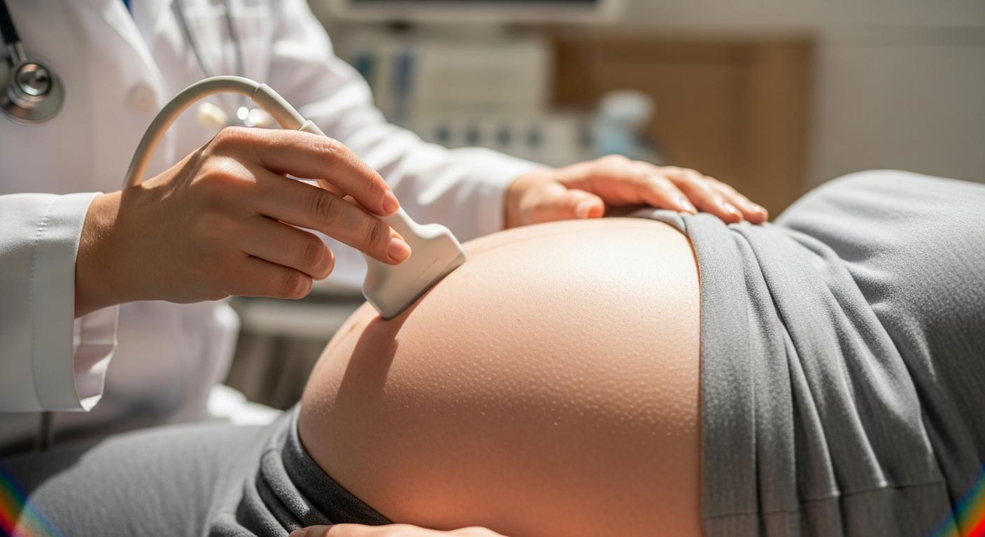
At the clinic, the session is led by a trained sonographer, obstetrician, or sometimes a midwife. You’ll recline on a couch, cool gel smoothed over your belly as the probe creates streams of images. While 2D imaging is the mainstay — capturing sharp, clinical measurements — 3D or 4D may be used for surface visuals when the baby is in an ideal spot, although all medical decisions are rooted in those 2D shots and Doppler flows for technical reliability.

Expect about 20–30 minutes for a single baby; slightly longer if twins or an elusive position challenge the clarity. Here’s a snapshot of steps you might experience:

  • Brief outline of what will be checked.
  • Systematic sweeps across the uterus, capturing biparietal diameter, head and abdominal circumference, femur length, all plotted on gestational age charts for growth trajectory.
  • Placental review: position (anterior, posterior, fundal), appearance, cord insertion.
  • Amniotic fluid: measured with precision — oligohydramnios (<5 cm or pocket <2 cm) and polyhydramnios (>24–25 cm or pocket >8 cm) flagged for action.
  • Confirmation of baby’s lie and their presentation.
  • Doppler flows: umbilical artery, sometimes uterine and middle cerebral arteries, if needed.
  • Summary feedback, immediate or in a detailed report.

Wondering about safety? Parent voices sometimes hesitate — sound waves only, no X-rays involved. The teams follow ALARA principles (minimum energy, optimal results). “Entertainment scans” without clinical oversight, however, are discouraged — it’s not a photoshoot, but a powerful medical tool.

What the Third Trimester Ultrasound Really Assesses

Fetal Growth: Are Percentiles Just Numbers?

Those growth charts and percentile lines can seem cryptic. In a third trimester ultrasound, several measurements — biparietal diameter (BPD), head circumference (HC), abdominal circumference (AC), femur length (FL) — all coalesce into an estimated fetal weight (EFW). However, here’s the clincher: EFW comes with an average margin of error, often ±10–15%. A baby that looks “big” or “small” in numbers may — on birth — surprise everyone! Interpretation always happens in context: parental build, previous scans, interval change, and any clinical red flags (for instance, maternal high blood pressure, diabetes, or history of small-for-dates baby) set the scene for decisions.

Challenges to accuracy? Perhaps baby’s head is deep in the pelvis, or their back hugs your front. Maybe prior C-section scars, high or low amniotic fluid, or body type affect how clear the picture comes out. Operator experience and machine quality count too.

Assessing Fetal Well-being and the Biophysical Profile

Sometimes, a deeper reassurance is needed. Here, the biophysical profile (BPP) may be used — watching not just movements, but the baby’s muscle tone, breathing-like motions, and heart activity. High scores suggest everything is ticking along well; borderline ones vote for closer follow-up, and if scores drop, it may prompt an urgent re-think: extra monitoring, possible supplementation for lung maturity (such as steroids), and discussions about timing birth.

Amniotic Fluid: Not Just About Volume

Fluid assessment is essential — too little (oligohydramnios) could hint at placental issues or a ruptured membrane, too much (polyhydramnios) sometimes signals diabetes or concerns with swallowing or gut function. Both extremes flag up for more frequent testing, tweaks in monitoring, and sometimes planning induction or C-section.

Placenta and Cord: Position and Function Matter

Is the placenta low (placenta previa), covering or too close to the cervix? Persistent issues close to term often mean planning a caesarean in advance for safety. Rarely, concern grows over placenta accreta spectrum (where the placenta “sticks” too deeply) or odd cord insertions — prompt specialist teamwork leads here. Eternal question: Is a loop of cord around the baby’s neck dangerous? Usually not — a “nuchal cord” is common and rarely impacts a healthy delivery plan, though it’s recorded for reference.

Baby Position and Delivery Planning

At around 32 weeks, breech or oblique positioning is not unusual. Most babies do an acrobatic roll to head-down by 36–37 weeks, but if not, an external cephalic version (ECV) may be considered — a gentle but skilled maneuver to turn baby, if the conditions are right. Uterine shape, placental site, or scars can affect whether ECV is prudent.

Doppler: Blood Flow Patterns, Not Just Pretty Pictures

When indicated (often in cases of suspected growth restriction), Doppler ultrasound steps in, assessing:

  • Umbilical artery for placental resistance.
  • Middle cerebral artery for baby’s “brain sparing” response.
  • Uterine arteries for maternal-side flow.

Changes here — say, high umbilical resistance or reversed blood flow, or low indices in the brain artery — drive closer monitoring. Decisions here are technical, evidence-backed, yet tailored to each scenario.

Cervical Assessment: When and Why

A transvaginal look at the cervix is reserved for when preterm labour risk is a query, rarely becoming routine after 28 weeks as prediction value decreases.

3D and 4D Imaging: More for Memories

While 3D/4D visuals can capture that first glimpse of a hand or yawn, the clinical backbone remains 2D. Sometimes, these images assist if facial or external anomalies are suspected, but their diagnostic value is limited compared to standard views.

Interpretation of Results and Birth Planning

So, what happens if the scan flags small-for-gestational age or suspected growth restriction? The third trimester ultrasound helps distinguish physiologically small babies from those at risk (for instance, due to placental issues), guiding whether more rigorous surveillance or even early delivery is considered. The flip side — suspected macrosomia — triggers a fresh debate on delivery route, risks of shoulder dystocia, induction, or planned caesarean.

Low or high fluid, placental changes, or persistent non-head-down positions? Each finding gets discussed dynamically, neither guaranteeing nor ruling out a specific birth path, but equipping parents and clinicians with real-time information.

Twins or multiples? The birth plan becomes even finer-tuned: chorionicity (do they share a placenta?), growth discrepancies, or unique Doppler patterns receive specialist attention.

Note, late scans rarely overrule your estimated due date unless absolutely no early dating exists to guide the process.

Special Situations and the Scan’s Limits

No ultrasound is omniscient. EFW gives an average estimate, but nature sometimes surprises everyone in the labour room. Technical issues — maternal body shape, fluid extremes, fetal position, even abdominal scars — may obscure clear pictures, sometimes triggering repeat scans or complementary tests.

Pregnancies complicated by diabetes, hypertension, preeclampsia, autoimmune disease, or kidney issues call out for a tailored plan and usually more frequent, targeted scans.

Ultrasounds provide valuable reassurance, but cannot rule out all subtle structural, metabolic, or genetic difficulties — nor can they tell you everything about how your baby will thrive post-birth.

Practical Aspects: Cost, Coverage, and Next Steps

For those with medical indications (like high blood pressure or fetal concerns), insurance often covers the third trimester ultrasound — but check specifics with your policy. For self-pay parents, request cost estimates, and ensure documentation of the reason for your scan.

Afterwards, interpretation happens as a dialogue. Sometimes a repeat scan, more frequent tests, or specialist referral follows. Occasionally, it means hospital admission for daily observation (for example, serious fetal growth restriction). Key clinical results combine with your symptoms, blood pressure, and baby’s kicks to build an ongoing, responsive plan.

Abnormal Findings: What Comes Next?

Should the scan detect something unexpected — abnormal Doppler, restricted growth, low fluid, or malpresentation — further tests are ordered with urgency that matches the finding. This might mean another third trimester ultrasound, continuous fetal monitoring, MRI, or echocardiography for heart anomalies. Timing balances medical urgency and thoughtful family planning.

Clear, supportive communication is your right. Request a written summary, compile questions, and never hesitate to seek a second opinion if complicated news arises. Paired with medical expertise, emotional support becomes vital, especially in complex situations.

Sometimes, lifestyle changes such as reduced work, no travel, or even inpatient stay are suggested for maximum safety. All these steps, and their rationale, should be documented in your maternity record — ensuring everyone involved in your care is on the same path.

Key Takeaways

  • Third trimester ultrasound is a targeted, late-pregnancy scan focusing on growth, amniotic fluid, placental health, presentation, and baby’s well-being.
  • Commonly performed at 31–33 weeks, with additional scans if there are special concerns: growth, fluid, twins, maternal conditions.
  • Measurements include biparietal diameter, head/abdominal circumference, femur, plus Doppler, all guiding clinical care.
  • Results inform the next steps: frequency of checks, possible interventions, and birth planning — but are interpreted together with your whole pregnancy history.
  • The tool is safe, using sound waves only, with “as low as reasonably achievable” energy to protect both parent and fetus.
  • Emotional and practical support, clear communication, and multi-specialist input help families approach labour and birth with confidence.
  • For continuous guidance and tailored health questionnaires, explore the free Heloa app, unlocking expert advice for every stage.

Questions Parents Ask

How many ultrasounds do I need in the third trimester?
Most low-risk pregnancies need no routine scan after 20 weeks, unless there’s a medical condition or concern. If your clinician wants to check growth, baby’s position, or fluid, expect a single third trimester ultrasound at 32–36 weeks. If problems show up (growth issues, diabetes, high blood pressure, twins), scans might repeat every 1–3 weeks. Scans are timed to match your needs, not a set calendar.

Will the scan tell the baby’s sex late in pregnancy?
Often yes — if the baby’s position and fluid offer a clear view. Sometimes, if the baby’s head is low in the pelvis or the legs are crossed, it’s tough, even with the best machines. Clinics usually concentrate on health checks, not just gender, so ask in advance if knowing is important.

If a concern is spotted, will I know immediately? What is the next step?
For most urgent or important findings, your provider shares a verbal summary in the appointment. A report with measurements and recommendations follows. If something serious is noticed, a repeat third trimester ultrasound, Doppler testing, or even referral to a specialist (maternal-fetal medicine, neonatology) may be arranged right away. Additional steps are based on urgency, gestational age, and your unique context.

What exactly will you look for in my scan?
Growth, fluid, placenta, and baby’s presentation are standard. If you have high blood pressure, diabetes, or previous complications, Doppler and more frequent scans might be included.

What questions should I bring to my scan appointment?

  • How is my baby’s growth compared with last scan?
  • What is the estimated weight, and how confident is that number?
  • Is the baby’s head-down yet? If not, do I qualify for turning the baby (ECV)?
  • Where is the placenta? Any issues with cord?
  • Amniotic fluid is normal or needs follow-up?
  • Should extra monitoring, a biophysical profile, or repeat ultrasound happen?
  • How will this update affect our birth plan?
  • What’s the expected next step if intervention is needed?

Are third trimester ultrasounds safe, especially with more scans?
Ultrasound — both transabdominal and, where necessary, transvaginal — is widely regarded as safe, with no ionising radiation. Medical teams use minimum energy and shortest time to get clear answers, following international standards.

What happens if scan results are uncertain or images unclear?
If baby’s position, low amniotic fluid, or body type hide images, a repeat third trimester ultrasound may be scheduled or an alternative test suggested. Your team adapts based on your needs, always prioritising safety and clarity.

For any step along the way, practical support and expert knowledge are never far — consult your healthcare provider, and if you desire personalised, ongoing tracking and answers, don’t hesitate to download the Heloa app any time.

Close-up on a pregnant woman's belly receiving the probe pass during the 3rd trimester ultrasound

Further reading :

Similar Posts

Waiting for the last stretch and wondering what a third trimester ultrasound will actually tell you, and whether it will change your plan for birth. You want clarity, not jargon. You want to know when to schedule it, what it checks, what numbers matter, and how those results shape next steps if something unexpected appears. That is exactly what follows, from timing and preparation to growth charts, fluid thresholds, Doppler patterns, and how teams translate images into smart decisions.

What it is and how it differs from earlier scans

A third trimester ultrasound is a focused late pregnancy check. Earlier scans concentrate on dating and full anatomy. This one pivots to growth over time, amniotic fluid, placental position and appearance, fetal presentation, and tests of wellbeing that inform delivery timing. Think management, not a second anatomy survey. You might ask, will it feel different from my mid pregnancy scan. The flow is similar, the targets are different.

You will hear the broader family of terms too, like obstetric ultrasound, fetal ultrasound, and ultrasound scan. Some centers call it a growth scan or simply late pregnancy sonography.

Why and when to have it

  • 28 to 32 weeks, set a late baseline for growth, amniotic fluid, and placenta, and catch early change.
  • Around 32 weeks, a sweet spot that leaves time to adjust surveillance or delivery plans.
  • 34 to 36 weeks, check presentation, fluid, and evolving findings.
  • 36 to 38 weeks, tighten birth planning, confirm head down or breech, reassess fluid and placenta.
  • 38 to 42 weeks, add scans or combined testing if there are post term concerns or new symptoms.

Not every pregnancy needs a routine third trimester ultrasound. It is most helpful when the result could steer care, for instance a size and date mismatch, reduced movements, bleeding, diabetes in pregnancy, maternal hypertension, twins, suspected growth problems, or placental concerns. High risk pregnancies may need serial scans every two to three weeks if fetal growth restriction (FGR) is suspected. When third trimester ultrasound shows steady growth, normal fluid, and a well positioned placenta, the plan often stays the same, which is a relief.

How the scan is done and safety

Preparation before the appointment

  • No special diet or medicines are required.
  • A full bladder is usually not needed this late.
  • Avoid lotions or oils on the abdomen for one to two days prior, they can interfere with sound transmission.
  • Wear clothing that lifts easily.
  • Bring your referral, prior reports, and any recent labs that might be relevant.

Arrive a few minutes early. A partner or support person is often welcome, with limits based on space and privacy.

Who performs it and the technology used

A trained sonographer, obstetrician, or midwife acquires images. Decision making relies on two dimensional imaging, and Doppler when indicated. Three dimensional and four dimensional views may be offered if conditions are favorable, but the medical read leans on measurements and blood flow studies, not souvenir clips.

Duration and typical steps

Most third trimester ultrasounds take 20 to 30 minutes, sometimes longer with twins or a head tucked deep in the pelvis. A typical flow:

  1. Brief overview of what will be checked.
  2. Gel on the abdomen and a sweep across the uterus.
  3. Head, abdomen, and femur measurements to compute estimated fetal weight (EFW).
  4. Review of placenta, umbilical cord, and amniotic fluid volume.
  5. Confirmation of fetal presentation and lie.
  6. Doppler assessment of placental circulation when relevant.
  7. A verbal summary followed by a written report.

Curious to follow along. Ask the clinician to point out the head, the stomach bubble, the spine, the placenta. It can be reassuring and memorable.

Safety and precautions

Ultrasound uses sound waves, not ionizing radiation. Teams follow ALARA principles, as low as reasonably achievable, to use the minimum energy needed to answer the clinical question. There are no usual contraindications in late pregnancy, including with abdominal scars or twins. Prolonged entertainment only scans without medical oversight should not replace diagnostic imaging.

What the scan evaluates

Fetal growth and biometry

Third trimester ultrasound focuses on growth trend, not a single snapshot. The sonographer measures the biparietal diameter (BPD), head circumference (HC), abdominal circumference (AC), and femur length (FL), then plots them on gestational age charts. Values between the tenth and ninetieth percentiles often fall in a physiological range, but context matters, parental size, prior growth curve, and time between scans all influence interpretation. Are you wondering what percentile really means. It is a ranking compared with babies of the same gestational age, not a grade for your baby.

Estimated fetal weight, methods and accuracy

EFW algorithms combine AC, HC, and FL, sometimes BPD. These formulas are validated, yet every estimate carries a typical error of about ten to fifteen percent. Accuracy can be reduced when the head is engaged, fluid is very low or very high, the abdominal wall is thick or scarred, or the baby keeps a tricky position. Third trimester ultrasound does not weigh a baby, it models weight from shape. That model still guides important decisions, especially when FGR or suspected large size are on the table.

  • EFW helps when weighing the balance of continued maturation against risks of staying inside if the placental environment shows strain.
  • Growth trends reassure when the curve stays steady, even if a single percentile number looks small.

If you like specifics, clinicians may also comment on fetal biometric measurements and report a fetal weight percentile.

Fetal wellbeing and the biophysical profile

Beyond measurements, clinicians watch movements, tone, breathing motions, and heart reactivity. A biophysical profile (BPP) combines ultrasound parameters, that is breathing motions, gross body movement, fetal tone, fluid, with heart rate reactivity. Some centers pair it with nonstress test (NST) or cardiotocography (CTG). High scores reassure. Borderline results prompt closer follow up. Low scores often trigger urgent evaluation or delivery planning depending on gestational age. You may hear terms like fetal breathing movements (FBM) or kick counts. Plainly put, the baby should flex, extend, and practice breathe, and the heart should respond to these bursts of activity. Parents can complement this with fetal movement counting (kick counts) at home.

Amniotic fluid assessment

Fluid can be measured by amniotic fluid index (AFI) or single deepest pocket (SDP). Thresholds used in many services:

  • oligohydramnios: AFI less than 5 cm or deepest pocket less than 2 cm
  • polyhydramnios: AFI above 24 to 25 cm or deepest pocket above 8 cm

Low fluid can hint at placental insufficiency or membrane rupture. High fluid can accompany gestational diabetes or fetal swallowing or intestinal issues. Both situations usually lead to closer checks and sometimes a change in delivery timing.

Placenta and umbilical cord

Reports document placental location, anterior, posterior, or fundal, and the distance from the cervix. Persistent placenta previa near term often requires planned cesarean. Suspicion of placenta accreta spectrum (PAS) prompts delivery planning in a center with surgical and neonatal support. Cord details may include cord insertion (velamentous) when visible. A nuchal loop around the neck is common and usually not harmful, but teams note it for awareness in labor. Some reports describe placental maturity, occasionally referred to as placental grading, placental maturity, or placental calcifications, which are descriptive and interpreted in context.

Fetal position and presentation

Third trimester ultrasound confirms fetal presentation and lie. Head down is cephalic presentation. Bottom first is breech presentation, which can be frank, complete, or footling. Sideways is transverse lie. Around 32 weeks many babies still rotate. If breech persists near 37 weeks, an external cephalic version, a hands on turn on the outside of the abdomen, may be offered when conditions are suitable.

Doppler and placental circulation

Doppler evaluates blood flow, a window into placental function. Common indices:

  • umbilical artery Doppler for placental resistance
  • middle cerebral artery Doppler for brain sparing adaptation
  • uterine artery Doppler for maternal placental perfusion
  • cerebroplacental ratio (CPR), the relationship of brain and placental flows
  • Select centers add ductus venosus Doppler in complex growth restriction

Patterns that raise concern include rising umbilical resistance, absent or reversed end diastolic flow, high uterine artery resistance with notching, and a marked fall in cerebral indices. These, especially alongside FGR or abnormal fluid, lead to tighter monitoring and careful timing of delivery. Color maps, that is color Doppler, can be used to map vessels and cord insertion.

Cervical assessment when relevant

If preterm labor is suspected, a transvaginal scan may measure cervical length. After 28 to 30 weeks its predictive value falls, so it is not routine for all.

3D and 4D imaging

Three dimensional and four dimensional imaging can be lovely keepsakes and occasionally help visualize surface anomalies. Diagnostic decisions rely on two dimensional imaging and Doppler. Quality depends on fetal position, fluid in front of the face, maternal body habitus, and where the placenta sits.

Interpreting results and impact on the birth plan

Growth concerns, small and large babies

  • FGR or small for gestational age, below the tenth percentile with slowed trajectory and or abnormal Doppler suggests placental insufficiency. Surveillance usually increases. If early delivery is likely, corticosteroids for lung maturity may be considered depending on gestational age. Third trimester ultrasound guides that call.
  • Suspected large baby, EFW is imperfect. Decisions weigh shoulder dystocia risk against induction or cesarean risks, with diabetes status, pelvic factors, and labor history in the equation.

Amniotic fluid and placenta findings

Oligohydramnios near term often leads to closer testing or planning delivery. Significant polyhydramnios may prompt symptom relief, for instance posture or sleep adjustments, and planned timing if risks rise. Persistent previa or suspected accreta changes timing, mode, and setting of delivery and usually involves a center with surgical and neonatal backup.

Position, external cephalic version, and delivery options

If breech persists near 37 weeks and the conditions are favorable, external cephalic version may be offered. If it is not attempted or does not work, options include planned cesarean or, in selected units with specific expertise, vaginal breech birth. Third trimester ultrasound helps check the placenta location and cord position before a version attempt.

Twins and multiples

Management depends on chorionicity, whether twins share a placenta, growth discordance, and twin specific Doppler patterns. Timing of delivery and monitoring intervals are tailored, often with specialist input.

Dating and due date

Late scans rarely change the estimated due date unless there was no reliable early dating. Clinicians synthesize history, early scans, and current findings to guide care.

Special situations and limitations

Accuracy limits and what ultrasound cannot do

EFW has a usual error of about ten to fifteen percent. Image quality can be limited by position, fluid, scarring, or BMI. Some anomalies are subtle or develop after mid pregnancy. Ultrasound cannot detect many genetic or metabolic conditions. A normal third trimester ultrasound greatly lowers, but does not eliminate, the chance of structural problems.

Maternal and uterine factors

Higher BMI, abdominal scars, fibroids, or very low fluid can reduce clarity. These may lead to repeat scans or complementary tests. If you have questions, ask what might improve views, such as a short walk, a different maternal position, or a brief return visit.

Maternal medical conditions and complex pregnancies

Conditions such as diabetes, high blood pressure or preeclampsia, autoimmune disease, renal disease, or a prior stillbirth shape surveillance and timing. Complex findings often prompt coordinated planning with maternal fetal medicine and neonatal teams. Third trimester ultrasound is central to that choreography.

Practical logistics, coverage, and follow up

Coverage and costs

When there is a medical indication, many insurers cover late pregnancy imaging, though specifics vary by plan and region. For self pay scans, ask for an estimate and ensure the indication is documented.

After the scan, follow up and coordination

Your clinician will explain whether you need a repeat third trimester ultrasound, additional Doppler, a BPP, or specialist referral. Increased surveillance may include:

  • More frequent ultrasounds for growth, Doppler, or fluid
  • Fetal heart rate monitoring with NST or CTG
  • Hospital admission when daily observation is needed, for example significant FGR or preeclampsia

Plans are built from multiple pieces, symptoms, blood pressure, kick counts, clinic assessments, and scan results. Together they support wise choices about induction or cesarean timing when needed. For reference, many services harmonize care with ACOG guidelines and NICE guidelines.

If an abnormality is found

Confirmatory tests and specialist referrals

Reasonable next steps include:

  • Repeat ultrasound or Doppler to confirm a trend rather than a one off variation
  • Fetal heart rate monitoring with NST or continuous CTG
  • Fetal MRI for brain or spine detail in selected cases
  • Fetal echocardiography if a heart concern is suspected
  • Referral to a specialized prenatal center and planning with neonatal teams

The sequence balances medical urgency with time to understand and plan.

Communication and emotional support

Ask for a clear written summary you can read later. Keep a small list of questions on your phone, it helps in the moment. Seeking a second opinion is reasonable, particularly for complex or rare findings. Perinatal psychological support can help you process uncertainty and decisions without feeling rushed.

Practical arrangements

Depending on the situation, your team may suggest work leave, less travel, or hospital admission to support a safer end of pregnancy. Key information is documented in your maternity record so everyone stays aligned on the plan for birth and newborn care.

Questions to ask your provider

  • How does my baby growth compare with prior scans, and what are the percentiles
  • What is the estimated fetal weight (EFW) and the confidence range
  • What is my baby presentation, and am I a candidate for external cephalic version if breech
  • Where is the placenta, how far is it from the cervix, and is the umbilical cord insertion normal
  • What is my amniotic fluid index (AFI) or single deepest pocket (SDP) and do I need follow up
  • Do I need umbilical artery Doppler, middle cerebral artery Doppler, uterine artery Doppler, or a biophysical profile (BPP) based on today findings
  • How do these results affect my timing and place of delivery
  • If an intervention is suggested, what are the next steps and who coordinates care

Putting it all together, practical scenarios

  • You have steady growth at the twentieth percentile, normal AFI, and normal Doppler. Third trimester ultrasound supports expectant management with routine visits and kick counts.
  • You have AC lagging, low AFI, and elevated umbilical resistance. Third trimester ultrasound prompts closer surveillance, steroid consideration depending on timing, and planning for earlier delivery if the pattern persists.
  • You have persistent breech at 37 weeks, a posterior placenta, normal fluid, and no contraindications. Third trimester ultrasound supports offering external cephalic version with appropriate monitoring.
  • You have twin pregnancy with growth discordance and abnormal CPR in one twin. Third trimester ultrasound guides a twin specific plan with specialist input and more frequent checks.

Why parents search for a third trimester ultrasound, common questions answered

  • Is it safe. Yes. Ultrasound uses sound waves, and teams follow ALARA to keep energy exposure low.
  • Will I get 3D pictures. Sometimes, if the position and fluid cooperate. The medical read relies on two dimensional views.
  • Can it change my due date. Rarely, unless no reliable early dating exists.
  • Can it predict my birth weight exactly. No. It estimates, and clinicians pair that estimate with your history and the baby growth trend to guide decisions.

A quick note on language. You will see third trimester ultrasound called a late pregnancy growth scan in some settings. Others will refer to it simply as obstetric ultrasound. These are different labels for the same practical goal, a clear look at growth, fluid, placenta, presentation, and circulation as you approach birth.

Key takeaways

  • A third trimester ultrasound focuses on growth, presentation, placenta, fluid, and wellbeing. It informs safe, flexible birth planning.
  • A common timing is 31 to 33 weeks, and there may be additional checks at 34 to 36 and 36 to 38 weeks if needed.
  • EFW has a typical error of about ten to fifteen percent. Trends and clinical context matter more than one number.
  • Fluid thresholds matter, AFI less than 5 cm or deepest pocket less than 2 cm suggests oligohydramnios, AFI above 24 to 25 cm or deepest pocket above 8 cm suggests polyhydramnios.
  • Doppler of the umbilical, uterine, and middle cerebral arteries can flag placental stress and guide timing.
  • Medically indicated ultrasounds are safe and follow ALARA. Keepsake scans should not replace diagnostic imaging.
  • Clear communication, coordinated planning, and compassionate support help parents make confident decisions.
  • For tailored advice and handy health tools, download the application Heloa for personalized tips and free child health questionnaires.

Third trimester ultrasound appears many times in this page because it is the anchor of late pregnancy surveillance, and because the right scan at the right time can turn anxiety into informed action. If you still have questions, bring them to your next visit and ask how your third trimester ultrasound will be used to shape a plan that fits your family.

Questions Parents Ask

How many ultrasounds will I need in the third trimester?

It depends on your pregnancy. Many low‑risk pregnancies need no routine late scan or only a single check around 32–37 weeks to confirm presentation and growth. If there are concerns—diabetes, high blood pressure, prior growth issues, twins, or any abnormal findings—scans may be repeated every 1–3 weeks or as directed by your care team. The frequency is chosen to balance reassurance with medical need: more scans when a trend must be watched closely, fewer when things are stable. If you feel anxious about timing, ask your provider what pattern they expect and how each scan would change care.

Can a third‑trimester ultrasound reliably tell me the baby’s sex?

Often yes, but not always. The baby’s position, low fluid, or fetal head engagement can hide the genital area late in pregnancy. When the anatomy is visible, sex identification is usually accurate, but visibility—not the machine—limits certainty. Remember some clinics focus only on medical checks and may not volunteer or prioritize sex determination; feel free to ask before the scan if you want to know and whether the sonographer can try to look.

If the scan shows a concern, how quickly will I be informed and what happens next?

You will usually get an immediate verbal summary at the appointment and a written report soon after. Urgent abnormalities are typically communicated the same day and trigger next steps like a repeat scan, Doppler studies, a biophysical profile or fetal monitoring (NST/CTG) within hours to days depending on severity and gestational age. Some situations lead to referral to a maternal‑fetal medicine specialist, fetal MRI, or discussion of earlier delivery; others mean closer outpatient surveillance. If you want clarity, ask at the scan who will call you, expected timelines for any follow‑up, and who to contact if you notice reduced fetal movements or new symptoms. This helps you feel supported and prepared while clinicians sort the best plan.

Close-up on a pregnant woman's belly receiving the probe pass during the 3rd trimester ultrasound

Further reading :

Similar Posts